Open Distal Femur and Segmental Tibia Fractures with Floating Knee Injury
Score and Comment on this Case
Clinical Details
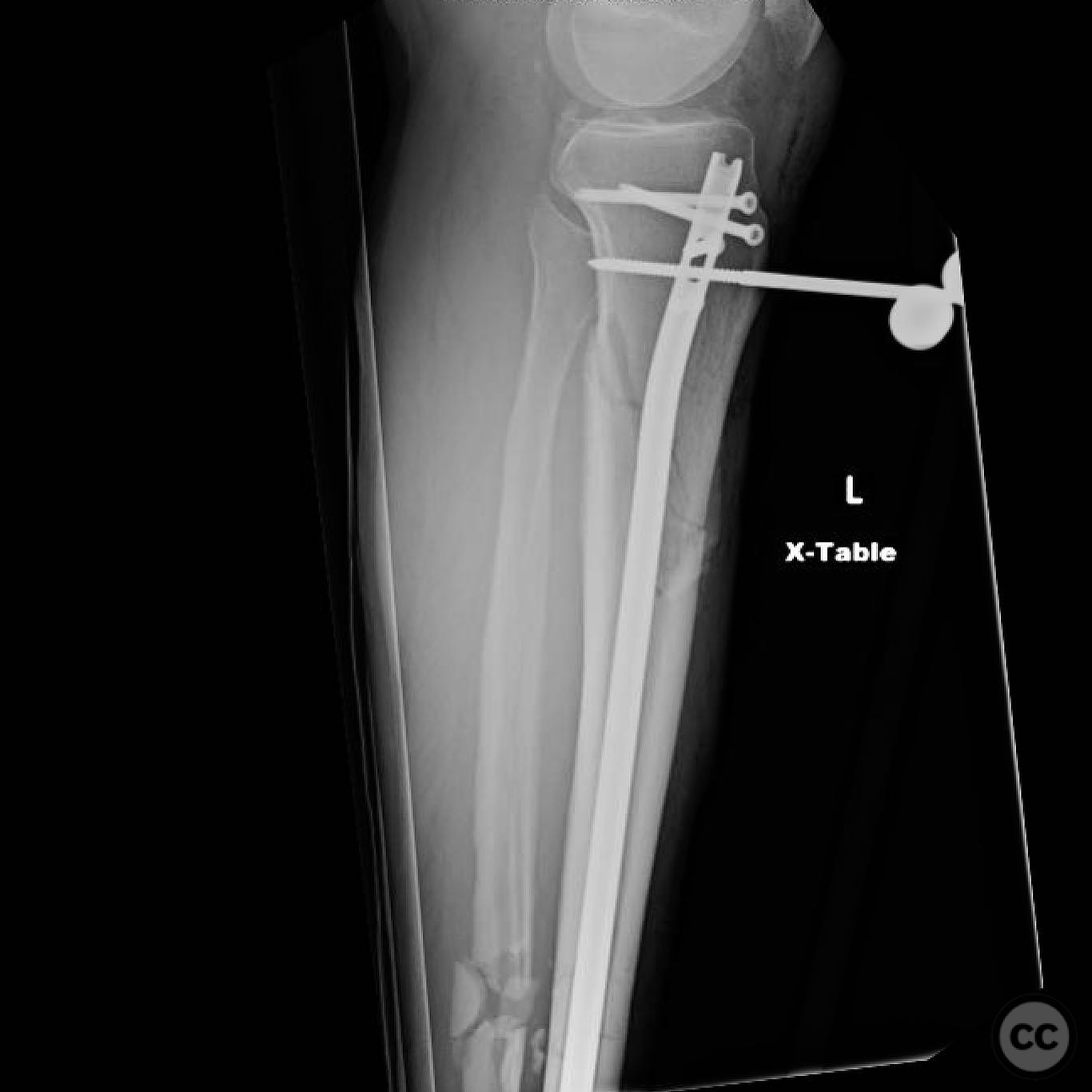
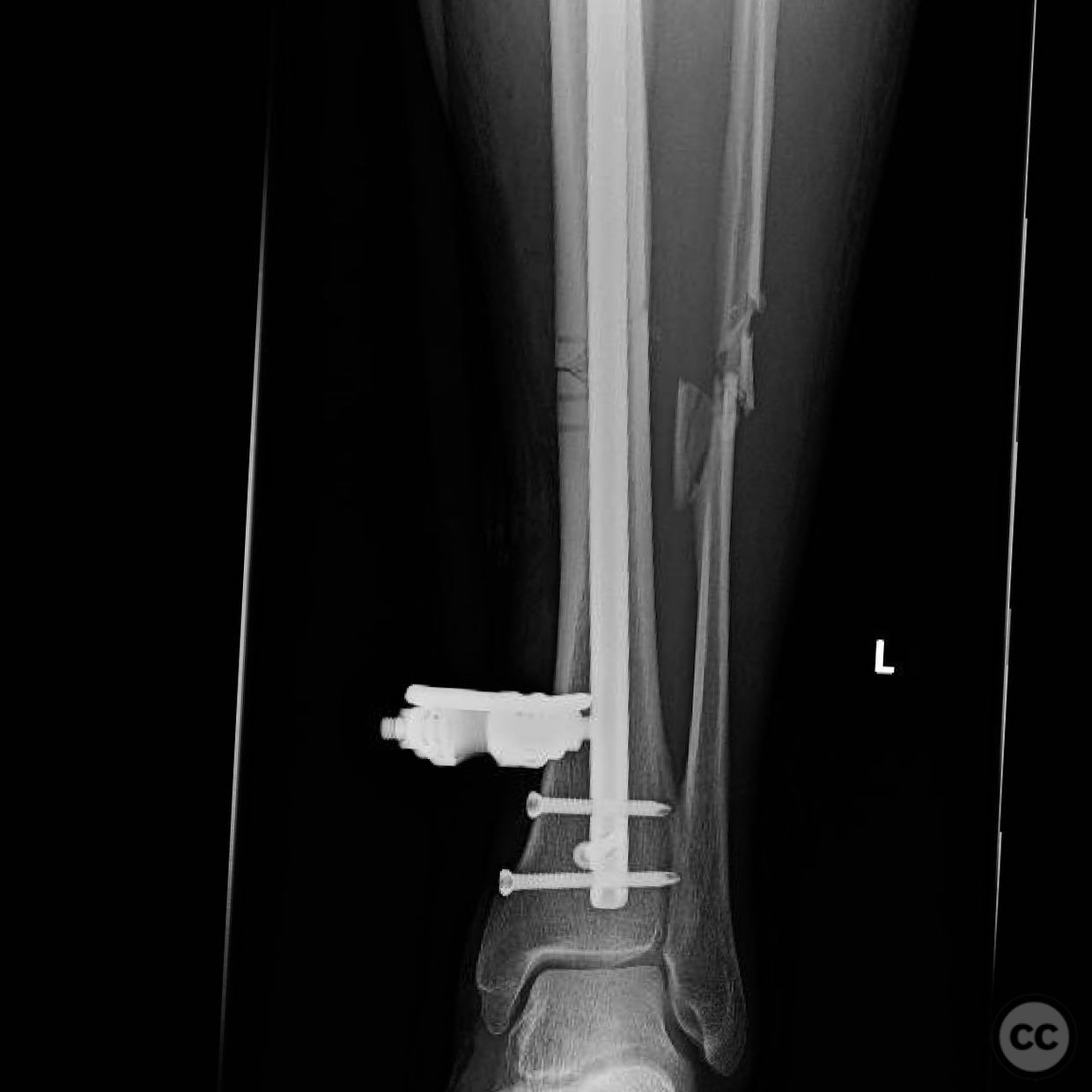
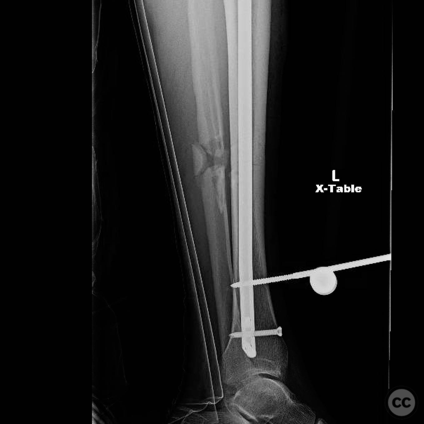
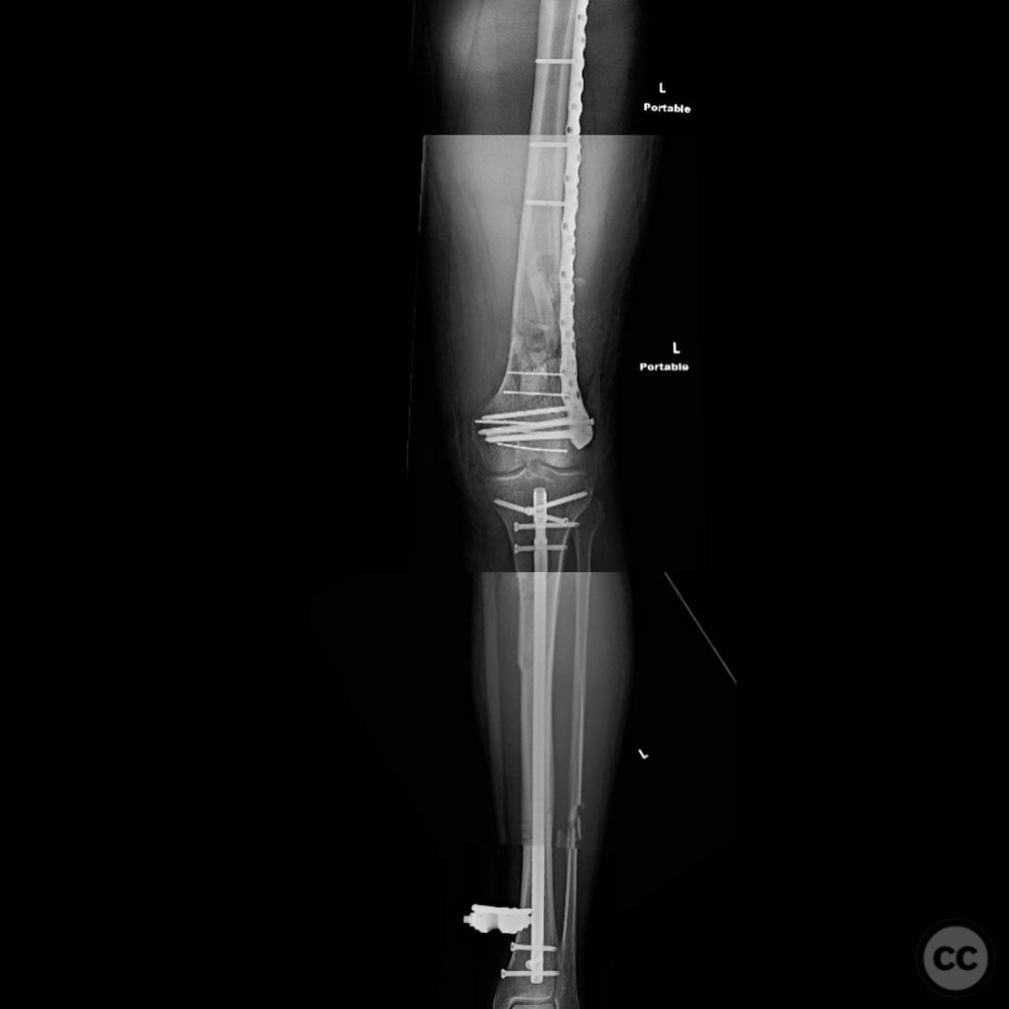
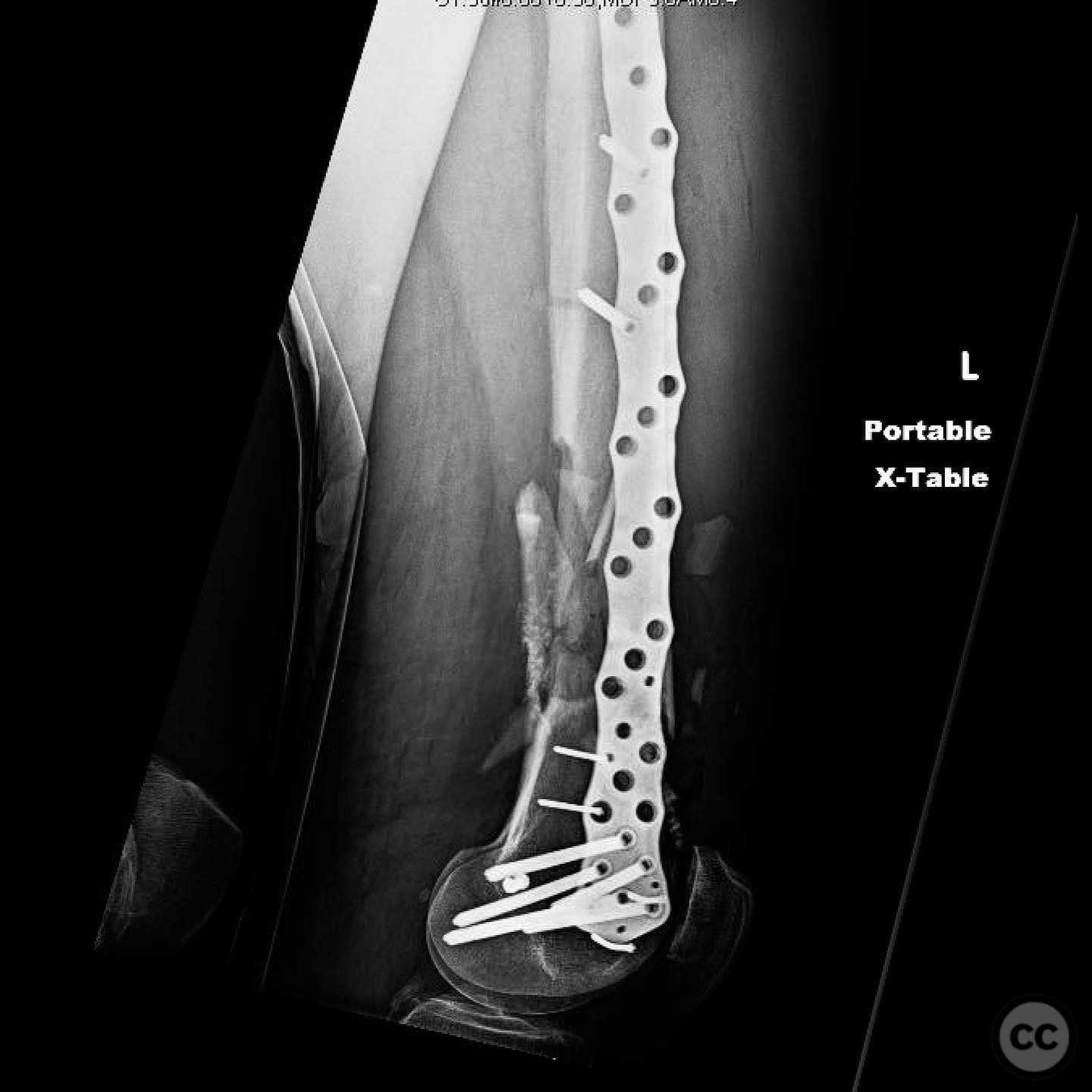
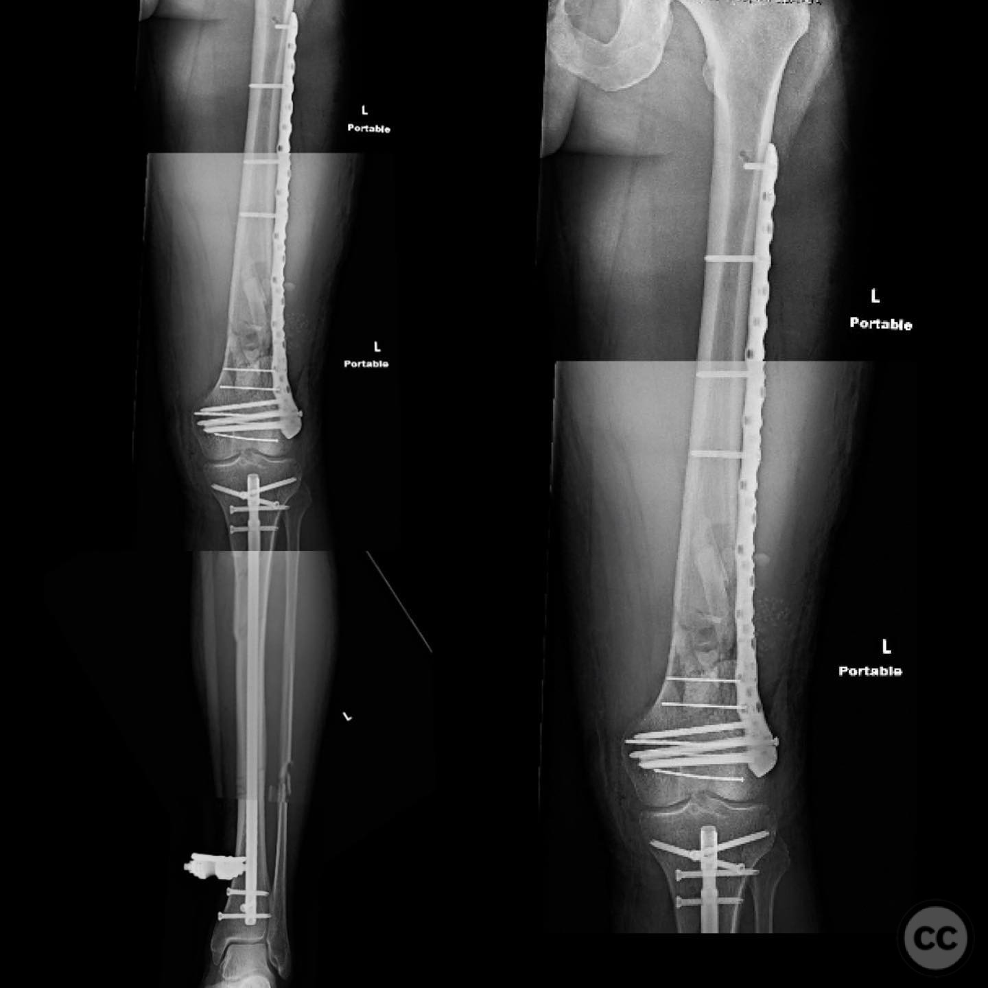
Clinical and radiological findings: A 44-year-old male cyclist was involved in a collision with a car, resulting in open fractures of the distal femur and segmental tibia. The femoral fracture presented with a 4 cm transverse wound on the anterior aspect, while the tibial fracture had a 2 cm anterolateral wound at the distal site. There were no associated head, chest, or abdominal injuries, and compartment syndrome was not present. Vascular examination was unremarkable.
Preoperative Plan
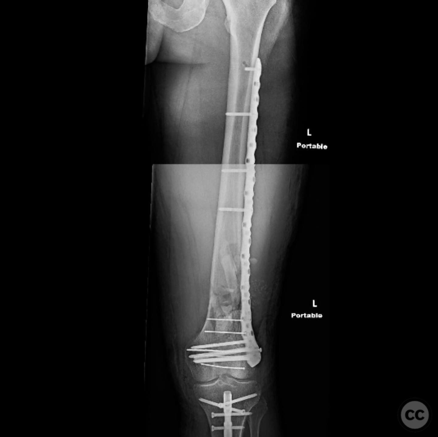
Planning remarks: The preoperative plan involved initial debridement and irrigation of both open fractures. The tibia was prioritized for intramedullary nailing, followed by spanning external fixation of the distal femur. Definitive fixation of the distal femur was planned for two days post-initial stabilization.
Surgical Discussion
Patient positioning: Supine positioning on a radiolucent table to facilitate fluoroscopic imaging. The affected limb was prepared for access to both the femur and tibia.
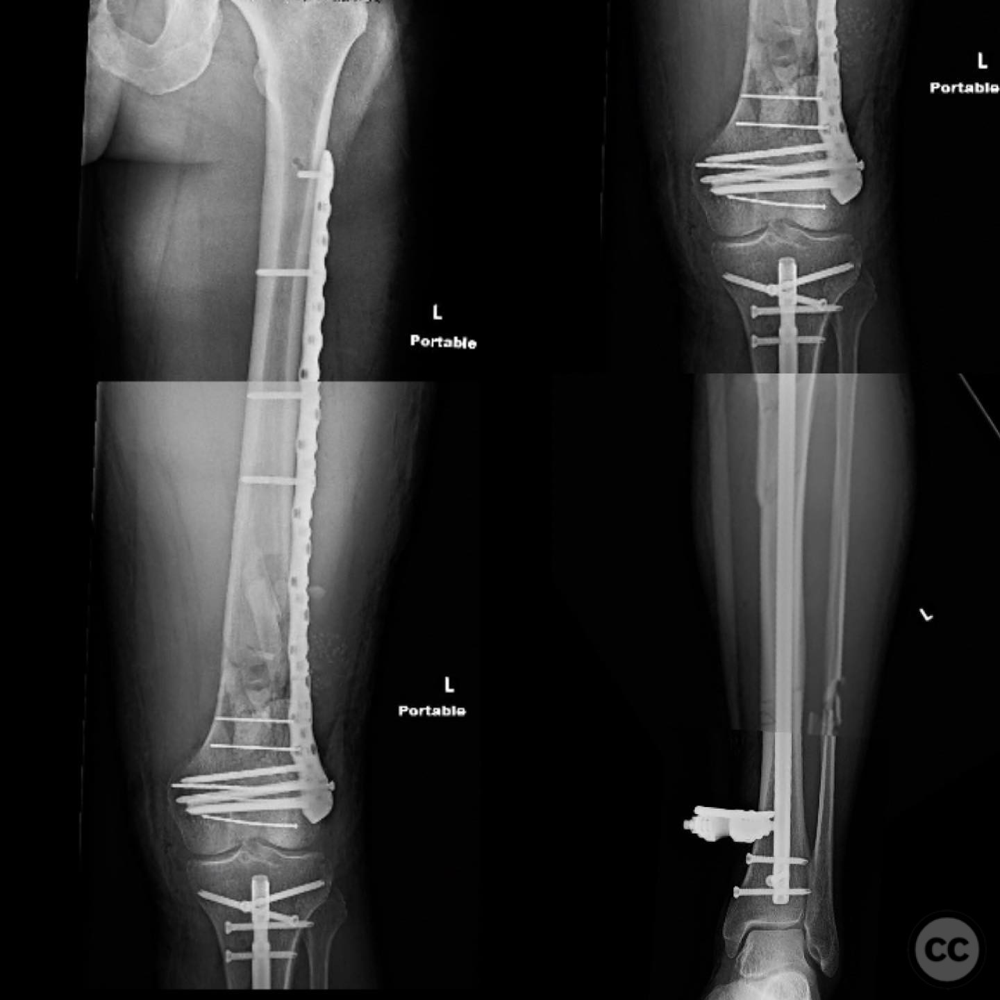
Anatomical surgical approach: For the tibia, a longitudinal incision was made to access the proximal tibial start site for intramedullary nailing. Percutaneous clamps were utilized for reduction, supplemented by blocking screws as needed. For the distal femur, an external fixator was applied initially, followed by an in situ lateral approach for plating after reduction was achieved.
Operative remarks:The tibial nailing required precise identification of the start site under fluoroscopic guidance, with percutaneous clamps aiding in reduction. Blocking screws were considered but not necessary due to successful reduction with clamps alone. The distal femur presented challenges in achieving coronal and sagittal alignment, necessitating reliance on the external fixator for reduction before proceeding with plating.
Postoperative protocol: Postoperative rehabilitation included early mobilization with weight-bearing as tolerated on the tibia, while the femur required protection until definitive fixation was achieved. Progressive weight-bearing was introduced following femoral plating.
Follow up: Not specified
Orthopaedic implants used: Intramedullary nail for tibia, external fixator for initial femoral stabilization, and locking plate for definitive femoral fixation.
Search for Related Literature

orthopaedic_trauma
- United States , Seattle
- Area of Specialty - General Trauma
- Position - Specialist Consultant

Industry Sponsership
contact us for advertising opportunities
Article viewed 288 times
14 Jul 2025
Add to Bookmarks
Full Citation
Cite this article:
Surname, Initial. (2025). Open Distal Femur and Segmental Tibia Fractures with Floating Knee Injury. Journal of Orthopaedic Surgery and Traumatology. Case Report 15666829 Published Online Jul 14 2025.