

Combined Anterior-Posterior Pelvic Ring Injury: Open Reduction and Internal Fixation with Posterior and Anterior Approaches
Score and Comment on this Case
Clinical Details
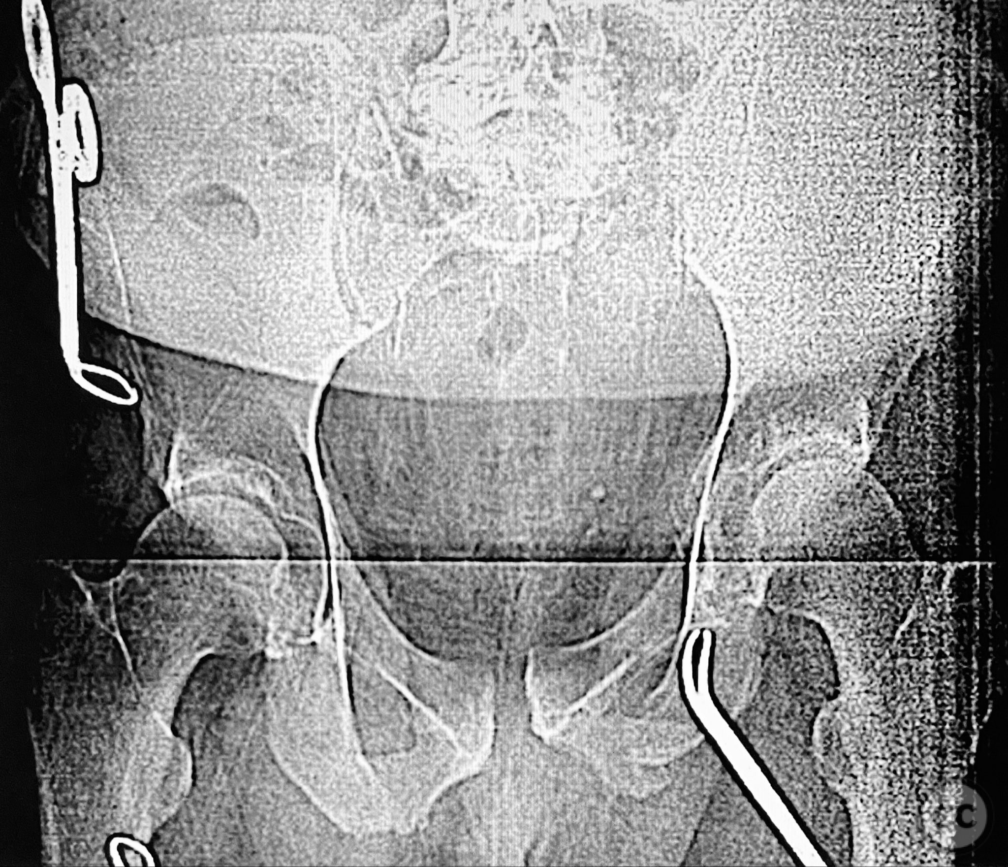
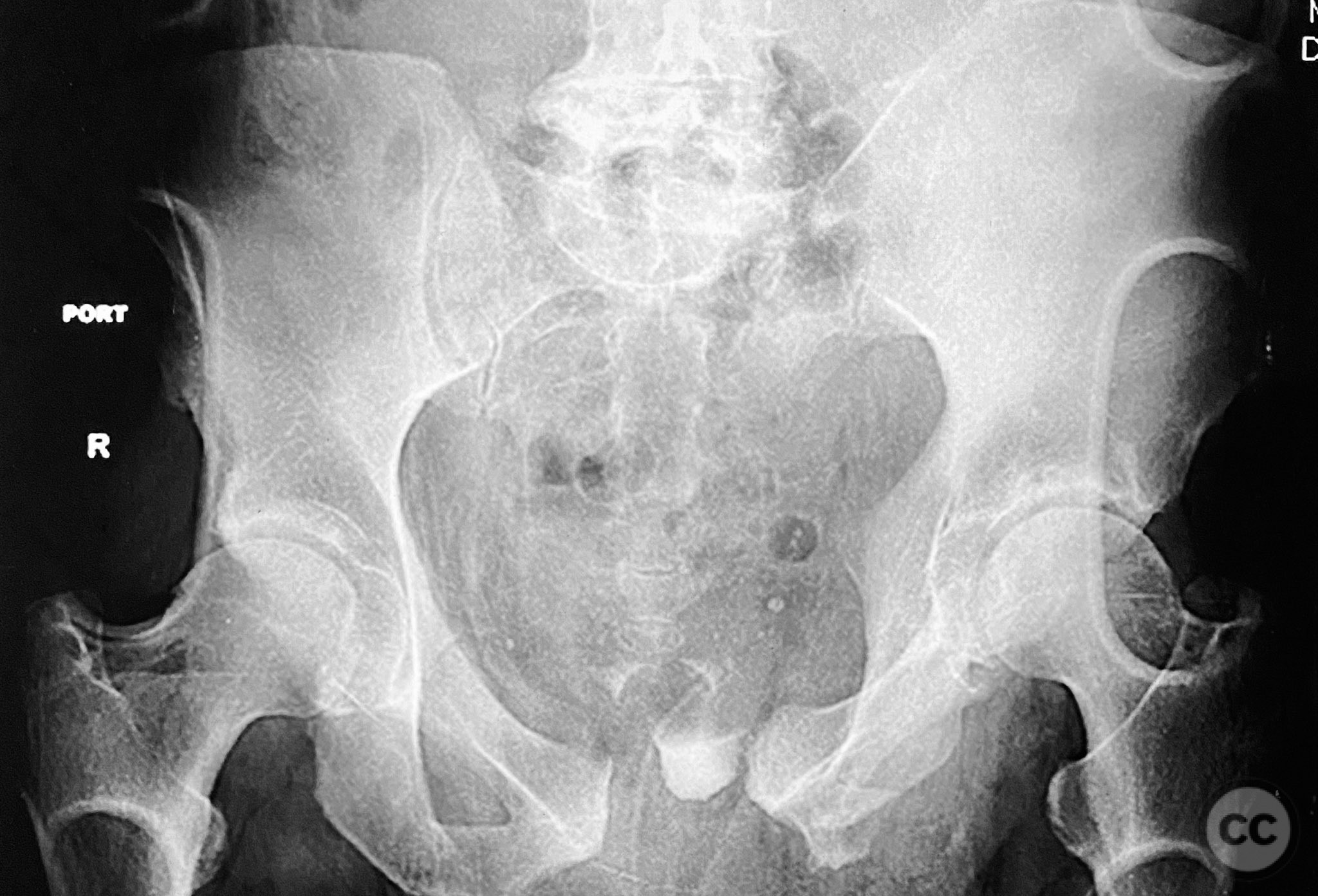
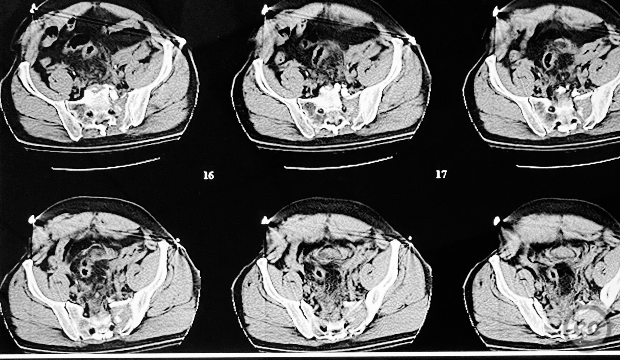
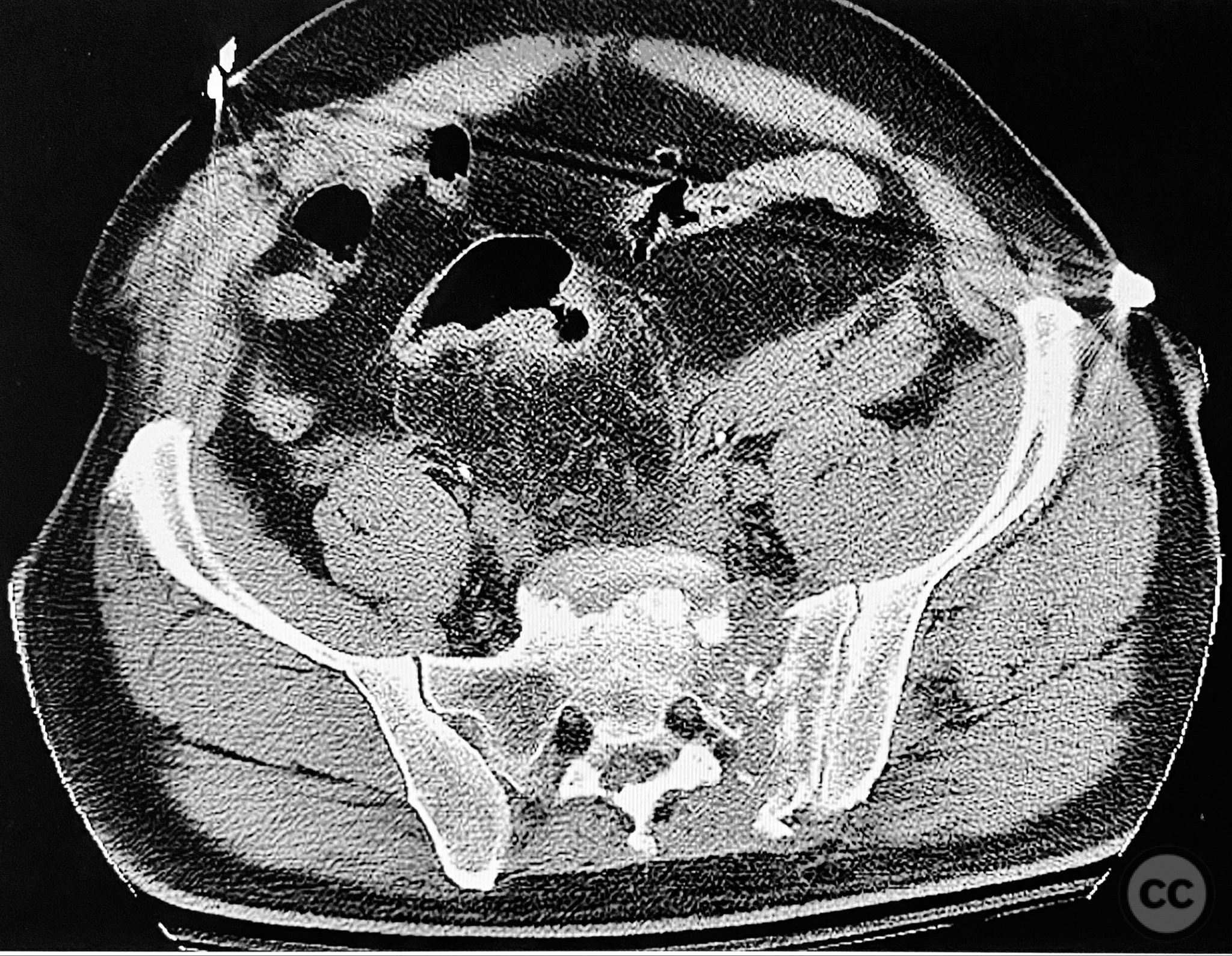
Clinical and radiological findings: A 54-year-old male presented following a motor vehicle accident with pelvic pain. He was haemodynamically stable and neurologically intact on admission. Initial anteroposterior pelvic radiograph demonstrated disruption of the symphysis pubis, fractures of the left superior/inferior pubic rami, and a left sacral fracture. Circumferential pelvic sheeting improved patient comfort and reduced hemipelvic displacement. Skeletal traction was considered but not yet applied. Computed tomography revealed comminution and displacement of the left sacral fracture, as well as detailed characterization of the anterior ring injuries. AO/OTA classification: 61-C1.3 (displaced, combined anterior and posterior ring injury with complete instability). Young-Burgess classification: APC III.
Preoperative Plan
Planning remarks: The preoperative plan included staged open reduction and internal fixation of the posterior pelvic ring via a dorsal approach in the prone position, utilizing direct visualization and reduction of the sacral fracture, followed by iliosacral and transiliac screw fixation. Definitive management of the anterior ring (symphysis pubis and ramus) was planned subsequently via an anterior approach in the supine position for reduction and stabilization.
Surgical Discussion
Patient positioning: For posterior fixation, the patient was positioned prone on a radiolucent table to facilitate direct dorsal access to the sacrum. For anterior fixation, the patient was repositioned supine.
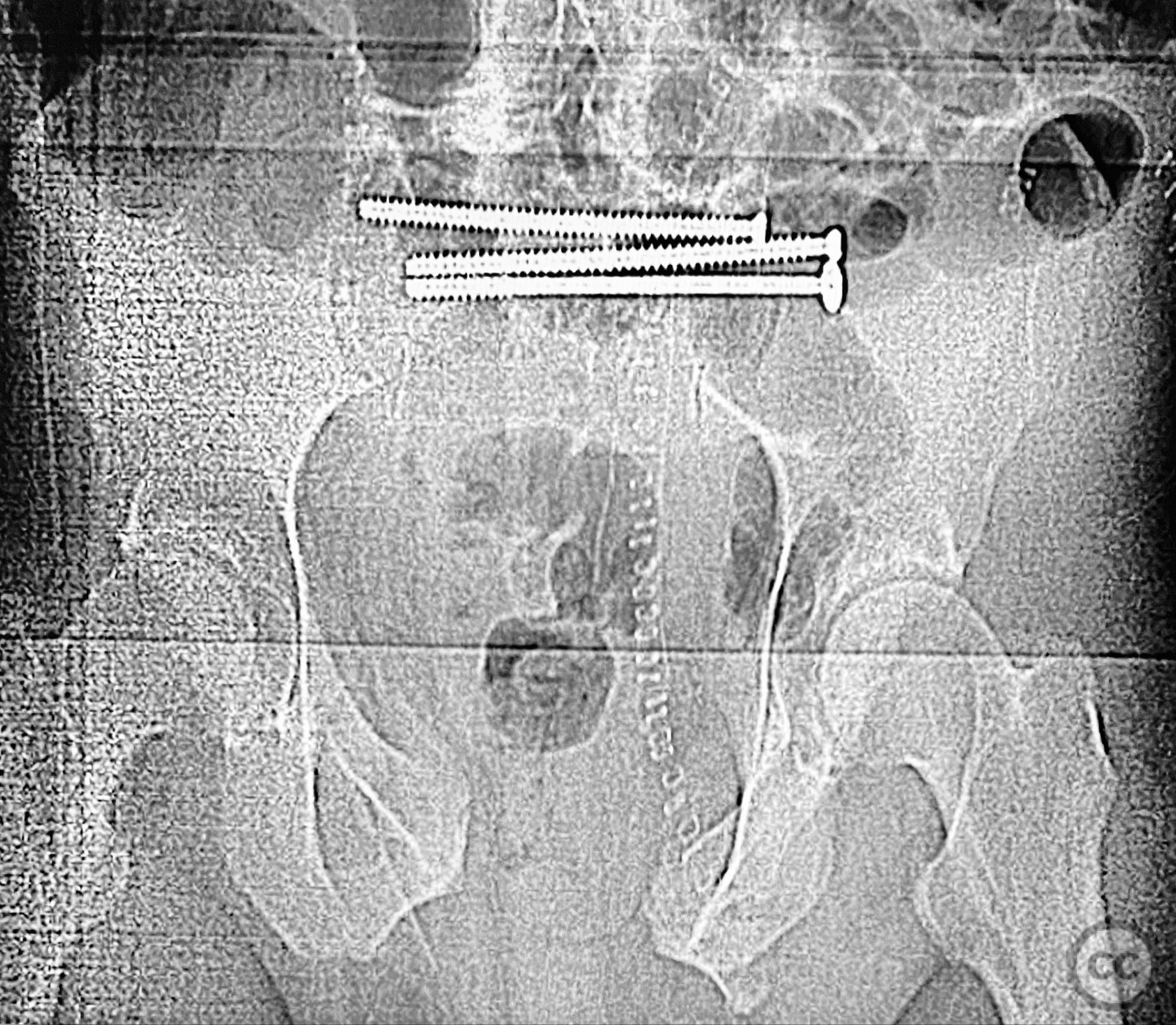
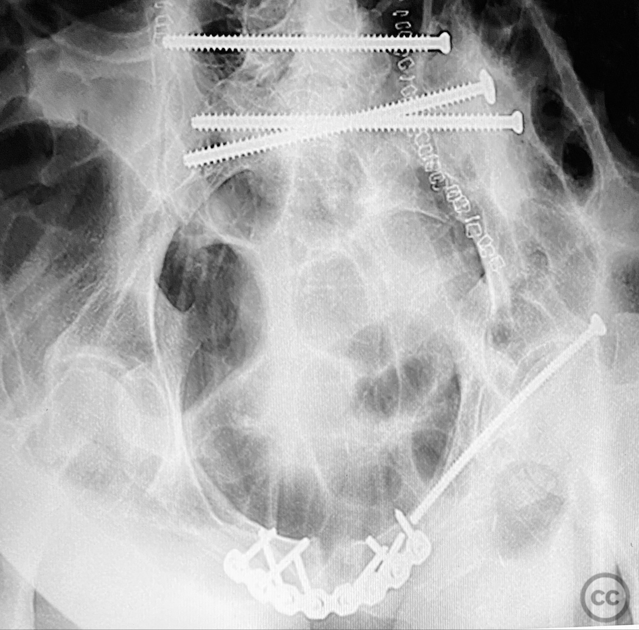
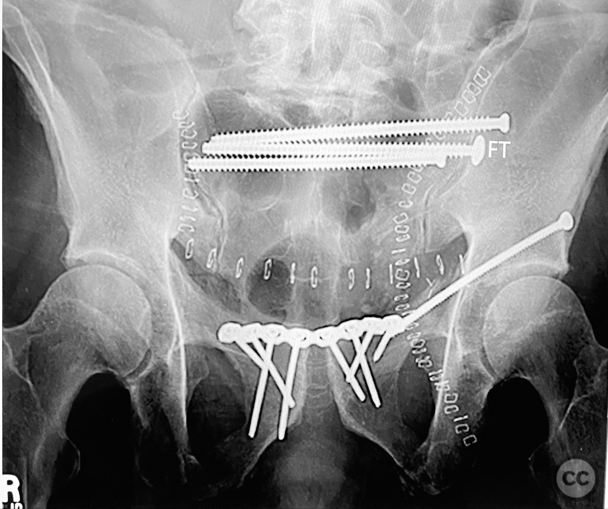
Anatomical surgical approach: The posterior approach involved a midline longitudinal incision over the sacrum, subperiosteal dissection to expose the dorsal sacral cortex, and direct assessment of fracture reduction using the cortical edges as reference points. Reduction was achieved and maintained with clamps. Iliosacral screws were inserted percutaneously under fluoroscopic guidance, supplemented by a posterior transiliac screw traversing from one ilium to the contralateral ilium across S1/S2. The anterior approach utilized a Pfannenstiel incision for exposure of the symphysis pubis and adjacent rami; reduction was achieved with pointed reduction forceps and stabilized with a symphyseal plate and additional ramus screws as indicated.
Operative remarks:The posterior reduction was facilitated by direct visualization of the dorsal sacral cortex, allowing precise assessment and clamping of the comminuted fragments prior to screw fixation. The iliosacral screws were placed without cannulated instrumentation longer than 130mm, reflecting historical implant limitations at the time of surgery. Postoperative CT confirmed satisfactory reduction and fixation of the posterior ring, though suboptimal positioning of iliosacral screws was noted (contained within the osseous fixation pathway but not optimized for maximal safety or precision). The anterior ring was subsequently addressed in a staged fashion, with anatomical reduction and stable fixation achieved via open technique. This case highlights technical challenges in achieving optimal screw trajectory and length, particularly with historical implant constraints, as well as the importance of staged management in complex pelvic ring disruptions.
Postoperative protocol: Early mobilization with protected weight bearing on the affected side for 8–12 weeks postoperatively. Thromboprophylaxis initiated perioperatively. Progressive advancement to full weight bearing as radiographic healing permits. Physiotherapy focused on core strengthening and gait training commenced within the first postoperative week.
Follow up: Not specified
Orthopaedic implants used: 7.3mm iliosacral screws (non-cannulated), posterior transiliac screw, anterior symphyseal plate, cortical screws for ramus fixation
Search for Related Literature


Industry Sponsership
contact us for advertising opportunities
Article viewed 406 times
12 Sep 2025
Add to Bookmarks
Full Citation
Cite this article:
Routt, ML. (2025). Combined Anterior-Posterior Pelvic Ring Injury: Open Reduction and Internal Fixation with Posterior and Anterior Approaches. Journal of Orthopaedic Surgery and Traumatology. Case Report 15634428 Published Online Sep 12 2025.