Associated Both Column Acetabulum Fracture with Keystone Fragment Involvement
Score and Comment on this Case
Clinical Details
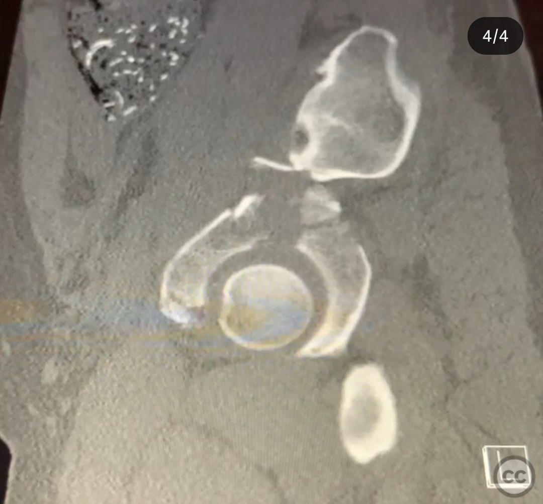
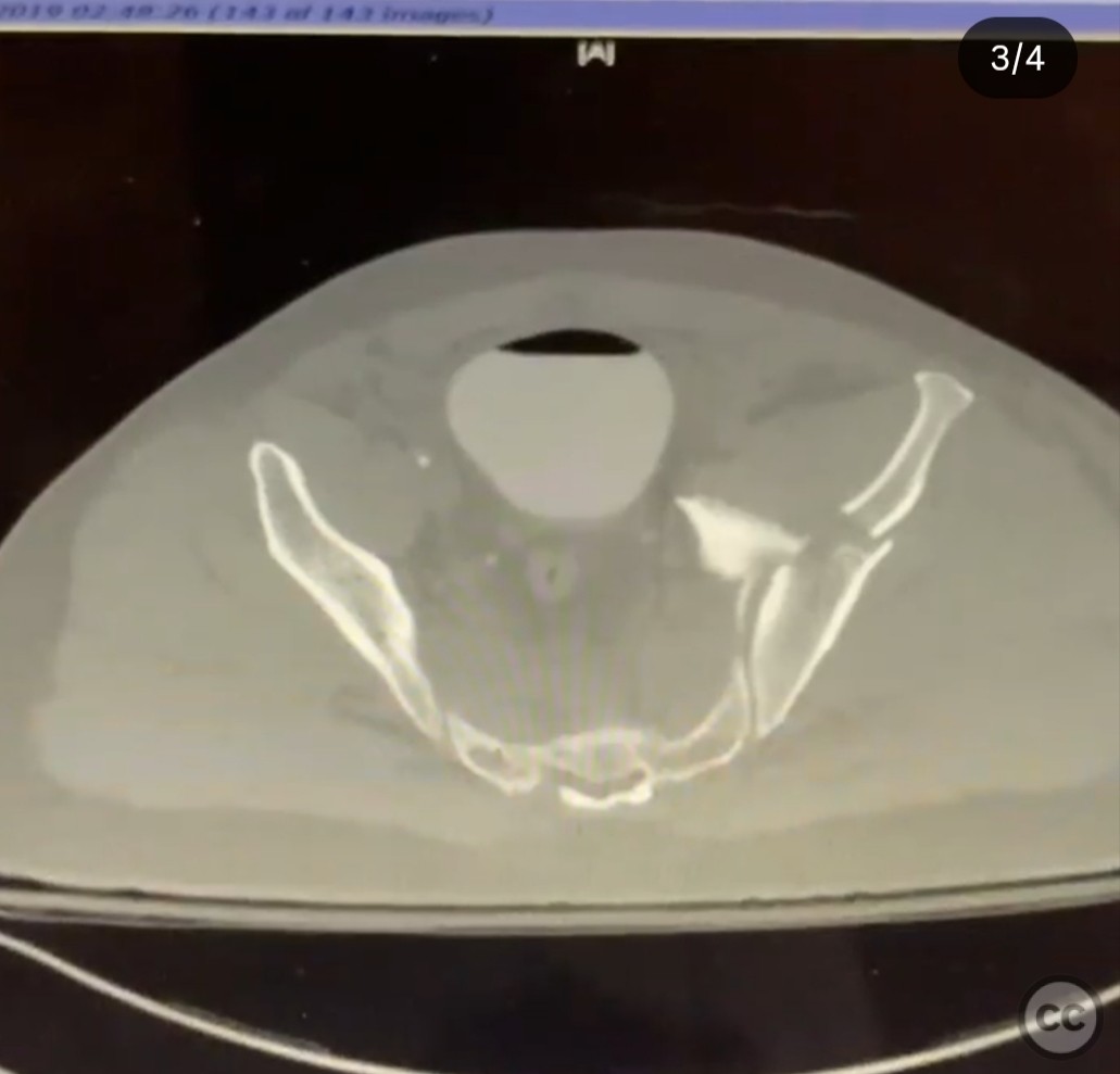
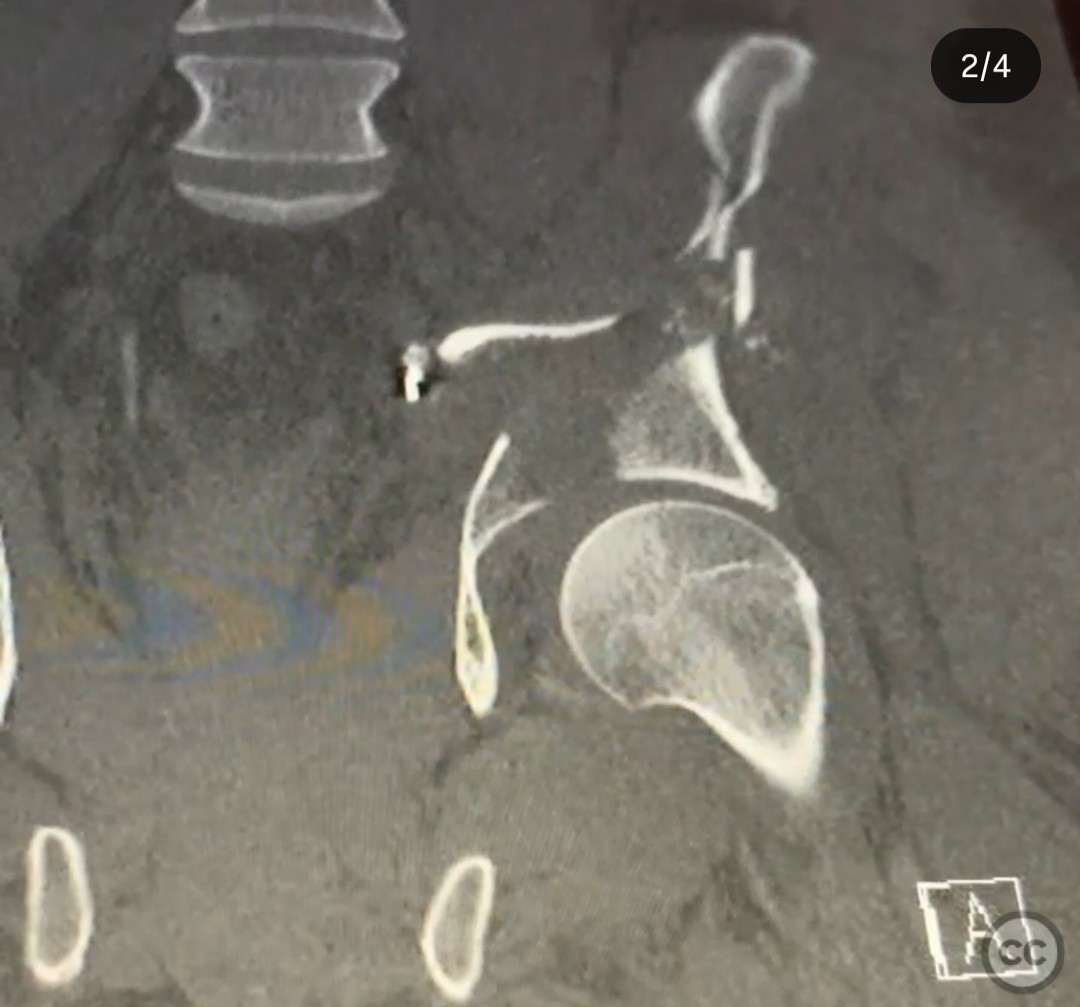
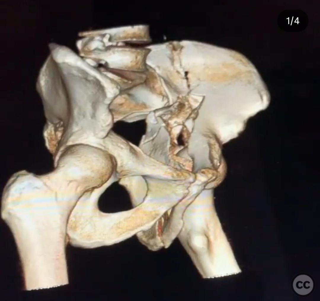
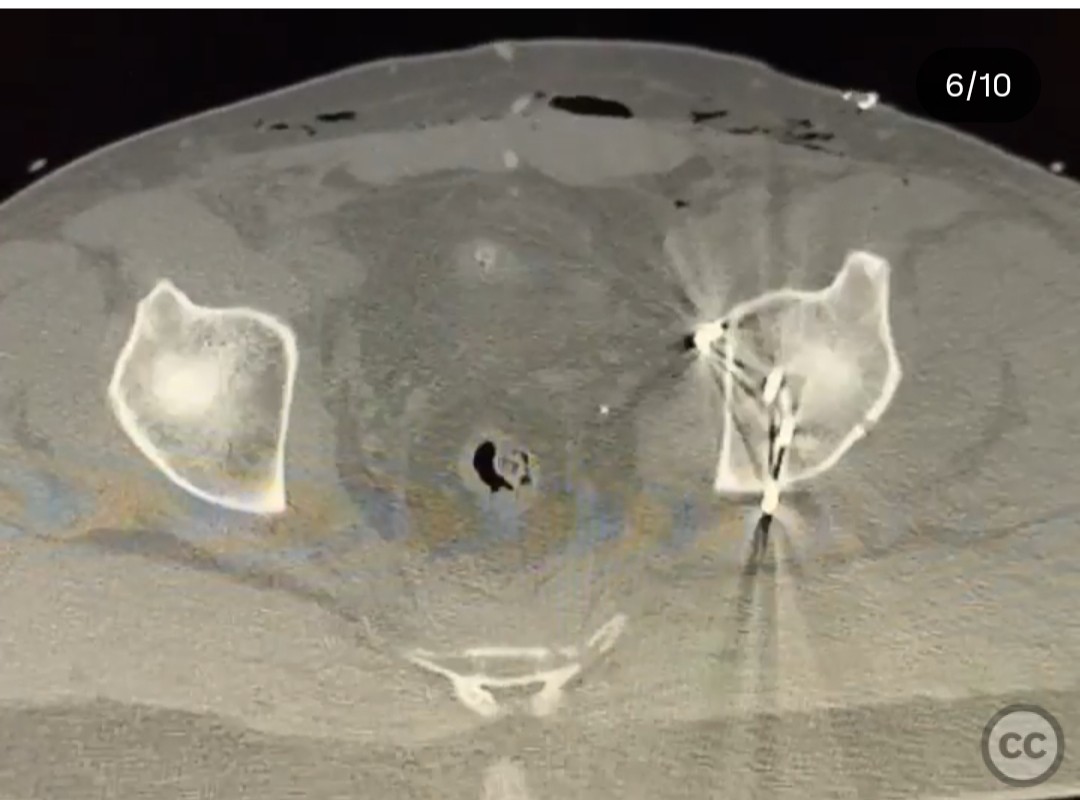
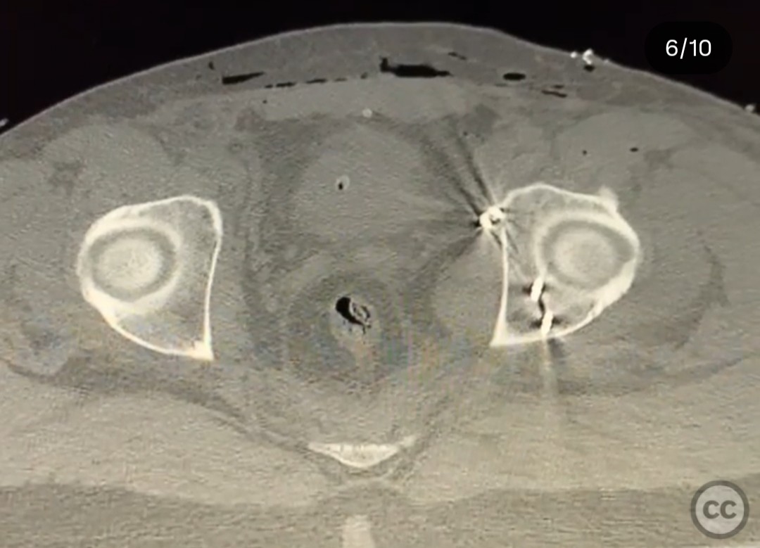
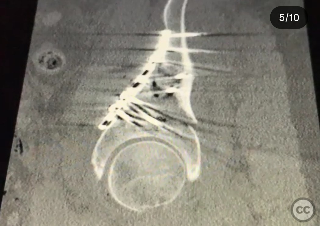
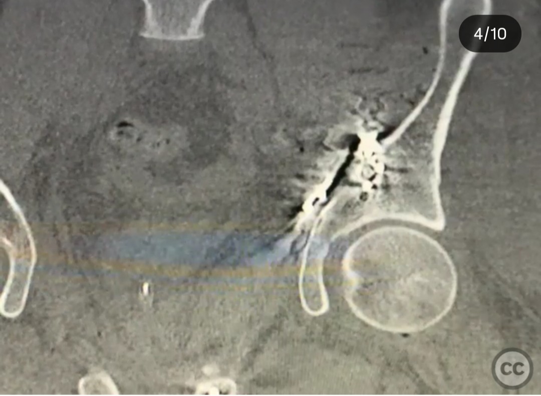
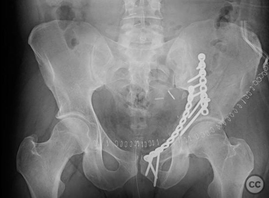
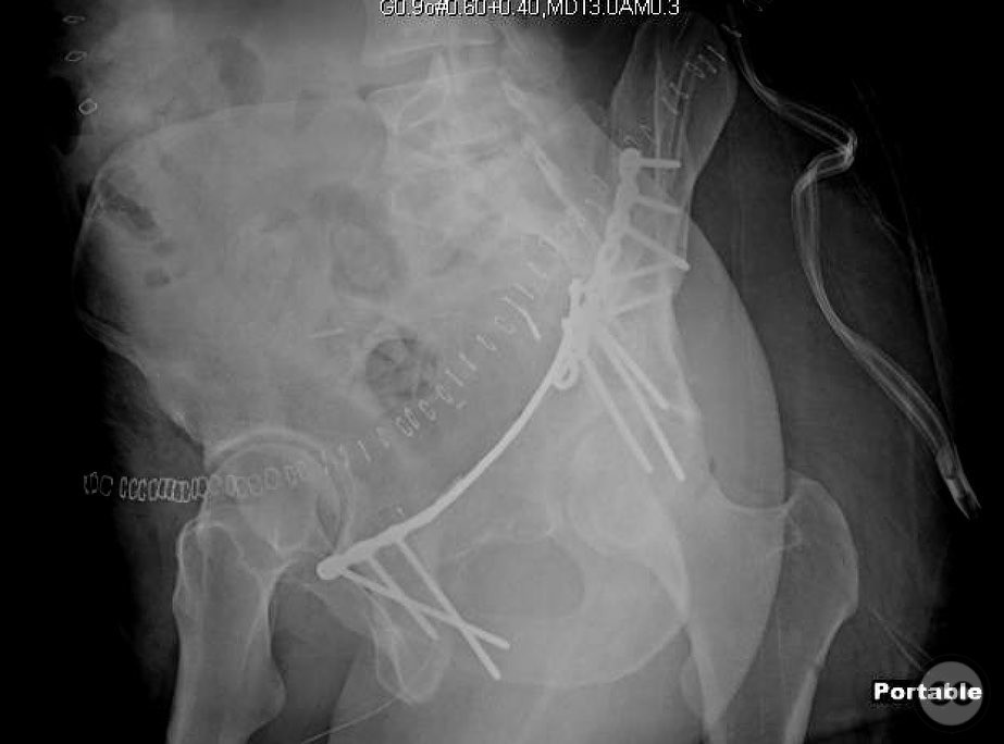
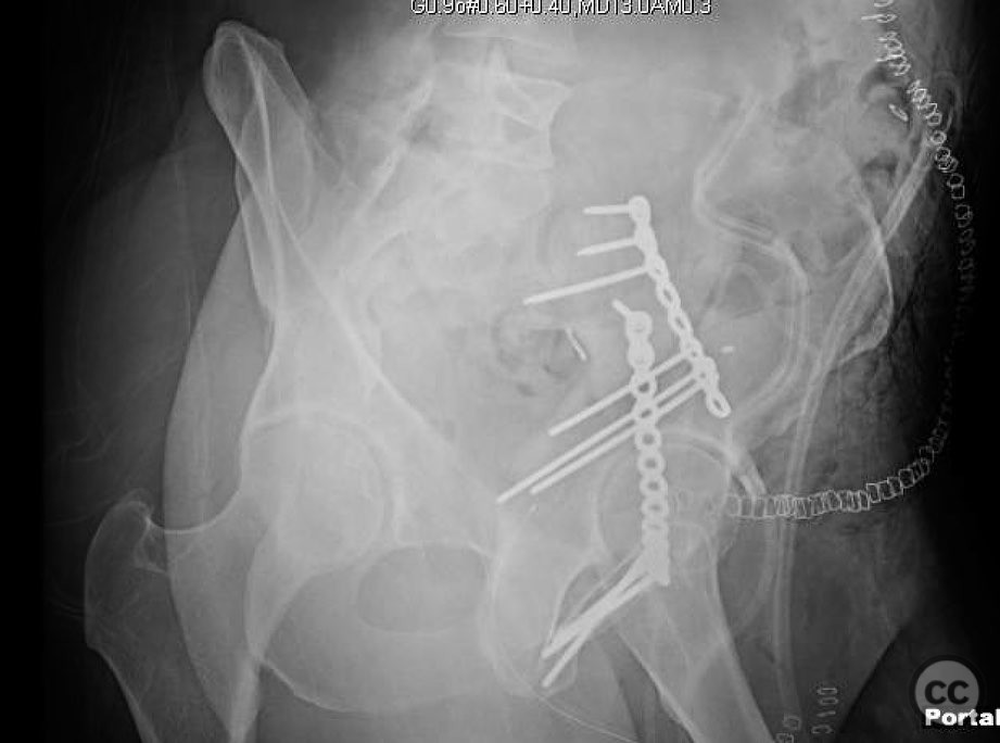
Clinical and radiological findings: A 26-year-old male sustained an associated both column acetabulum fracture while riding a motorcycle. Initial radiological assessment revealed a large keystone fragment nearly impaling the internal iliac vessels. The fracture involved both anterior and posterior columns, with a super posterior exit involving the sacroiliac joint. The patient was otherwise healthy with no additional injuries reported.
Preoperative Plan
Planning remarks: The preoperative plan involved an ilioinguinal approach to address the anterior column and keystone fragment. The reduction sequence was planned as follows: reduction of the anterior column exit at the iliac wing, anatomical reduction of the keystone fragment to the intact ilium, reduction of the anterior column at the pelvic brim or pubic ramus, and finally, reduction of the posterior column to the keystone and anterior column.
Surgical Discussion
Patient positioning: Supine position on a radiolucent table to facilitate intraoperative imaging and access to both anterior and posterior columns.
Anatomical surgical approach: A combined ilioinguinal and modified Stoppa approach was utilized. A midline Pfannenstiel incision was made to access the modified Stoppa window, allowing preperitoneal dissection to expose the quadrilateral plate, medial wall, and posterior column from the internal pelvis. The bladder was retracted inferiorly, and the corona mortis vessels were identified and ligated to prevent bleeding. Next, an ilioinguinal approach was performed through a standard oblique incision parallel to the inguinal ligament. Dissection was carried through the external oblique aponeurosis. The femoral neurovascular bundle and iliopsoas muscle were mobilized to allow access to the pelvic brim and anterior column through the lateral and middle windows. Careful manipulation was required around the keystone fragment, which was closely related to the internal iliac vessels, necessitating meticulous dissection and protection.
Operative remarks:The surgeon noted that the anterior column required derotation using a spiked pusher, followed by clamping with a goose clamp and bulldog clamp. The posterior column was reduced and clamped with a goose clamp over the pelvic brim. Despite a wide inferior aspect, fixation was deemed stable without additional screws in the symphysis or sacroiliac joint. A postoperative CT scan confirmed adequate fixation of the posterior column with three screws from the anterior column plate.
Postoperative protocol: Postoperative rehabilitation included protected weight-bearing for 6 weeks, followed by gradual progression to full weight-bearing as tolerated. Range of motion exercises were initiated early to prevent joint stiffness.
Follow up: Not specified.
Orthopaedic implants used: Orthopaedic implants used included a reconstruction plate for the anterior column and three screws for posterior column fixation from the anterior approach.
Search for Related Literature

orthopaedic_trauma
- United States , Seattle
- Area of Specialty - General Trauma
- Position - Specialist Consultant

Industry Sponsership
contact us for advertising opportunities
Article viewed 266 times
19 Jul 2025
Add to Bookmarks
Full Citation
Cite this article:
Surname, Initial. (2025). Associated Both Column Acetabulum Fracture with Keystone Fragment Involvement. Journal of Orthopaedic Surgery and Traumatology. Case Report 15624884 Published Online Jul 19 2025.