Tibiotalar Arthrodesis with External Fixation Following Post-Traumatic Osteoarthritis and Previous Osteomyelitis
Score and Comment on this Case
Clinical Details
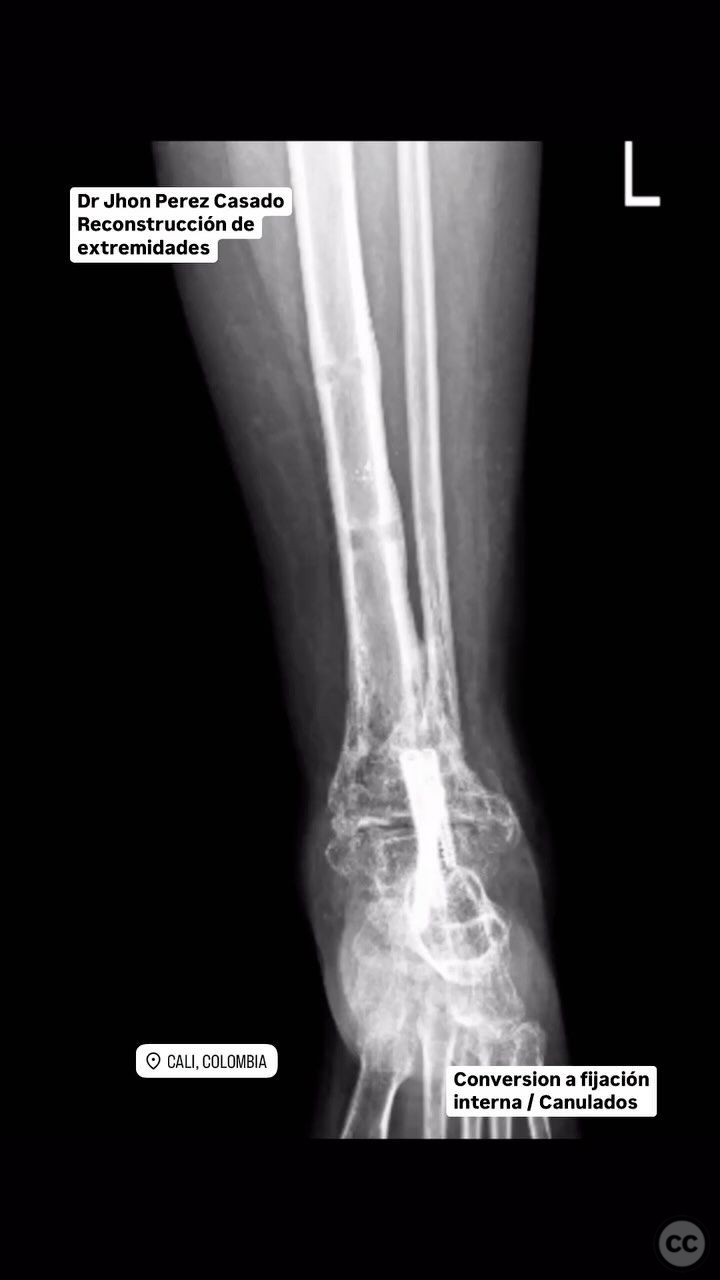
Clinical and radiological findings: The patient presented with post-traumatic osteoarthritis of the tibiotalar region, complicated by a history of previous osteomyelitis. Radiological assessment confirmed significant degenerative changes in the tibiotalar joint, consistent with post-traumatic osteoarthritis. There was no acute infection noted at the time of presentation.
Preoperative Plan
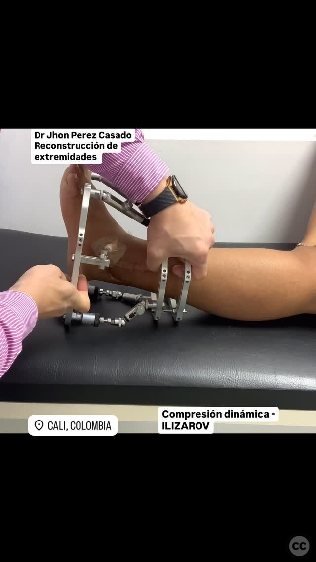
Planning remarks: The preoperative plan included bone resection to address deformity, followed by tibiotalar arthrodesis. The initial stabilization was planned using external fixation, with the potential for conversion to internal fixation using cannulated screws if necessary. Dynamic compression was considered to enhance arthrodesis.
Surgical Discussion
Patient positioning: The patient was positioned supine on the operating table, with the affected limb elevated and supported to allow for optimal access to the tibiotalar joint.
Anatomical surgical approach: A longitudinal incision was made over the anterior aspect of the ankle joint. Subcutaneous tissues were dissected to expose the tibiotalar joint. Careful debridement of osteoarthritic and necrotic bone was performed. The joint surfaces were prepared for arthrodesis, ensuring adequate bone contact for fusion. An external fixator was applied to maintain alignment and compression across the arthrodesis site.
Operative remarks:The surgeon noted that the previous osteomyelitis had resulted in some bone loss, which required meticulous debridement and preparation of the joint surfaces to ensure successful fusion. Dynamic compression was applied through the external fixator to promote bone healing. Conversion from external to internal fixation with cannulated screws was planned once initial stability and early healing were confirmed.
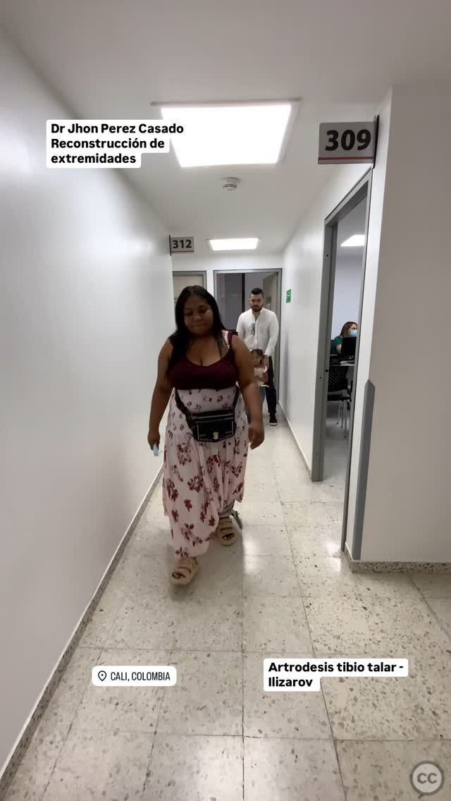
Postoperative protocol: Postoperatively, the patient was allowed full weight-bearing as tolerated with the external fixator in place. On-the-go training was encouraged to maintain mobility and muscle strength. Transition to internal fixation was planned once sufficient healing was observed.
Follow up: Not specified.
Orthopaedic implants used: External fixator, cannulated screws (for planned conversion).
Search for Related Literature

Jhon Perez Casado
- Colombia , Cali - Valle del Cauca
- Area of Specialty - Lower Limb
- Position - Specialist Consultant

Industry Sponsership
contact us for advertising opportunities
Article viewed 283 times
23 Jul 2025
Add to Bookmarks
Full Citation
Cite this article:
Perez Casado, J.J.. (2025). Tibiotalar Arthrodesis with External Fixation Following Post-Traumatic Osteoarthritis and Previous Osteomyelitis. Journal of Orthopaedic Surgery and Traumatology. Case Report 14218619 Published Online Jul 23 2025.