 Anatomic reduction (or as my partner says_ RONA- restoration of native anatomy). For _1.jpg)
 Anatomic reduction (or as my partner says_ RONA- restoration of native anatomy). For _2.jpg)
 Anatomic reduction (or as my partner says_ RONA- restoration of native anatomy). For _3.jpg)
 Anatomic reduction (or as my partner says_ RONA- restoration of native anatomy). For _5.jpg)
 Anatomic reduction (or as my partner says_ RONA- restoration of native anatomy). For _4.jpg)
 Anatomic reduction (or as my partner says_ RONA- restoration of native anatomy). For t(.jpg)
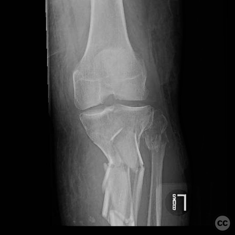
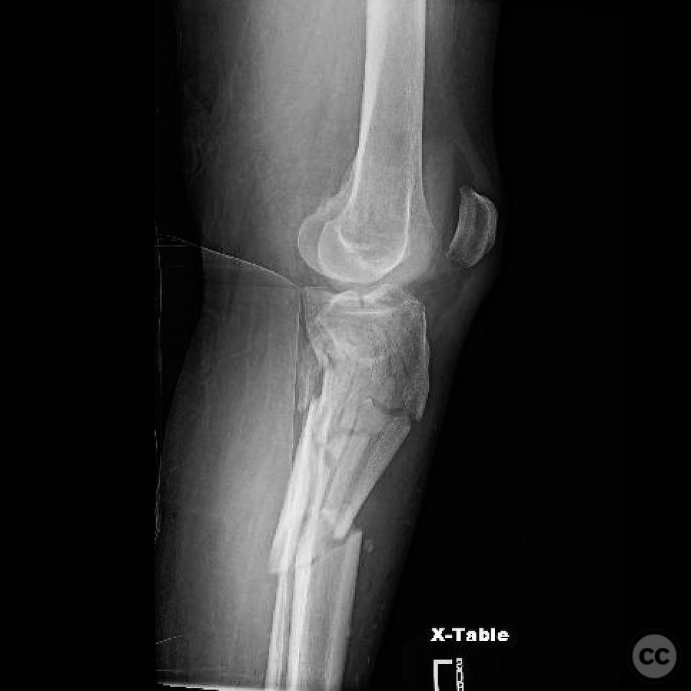
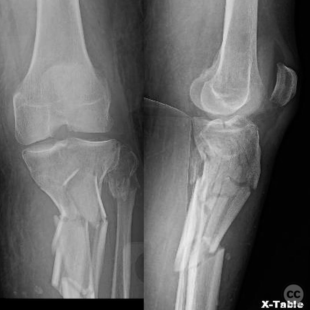
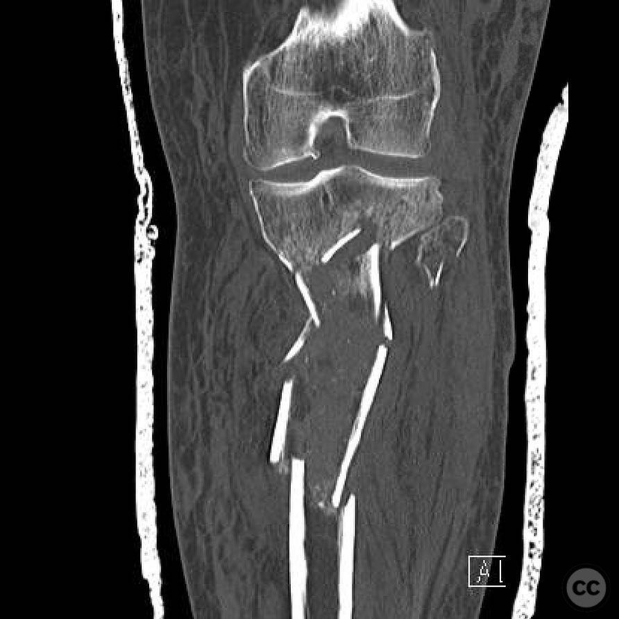
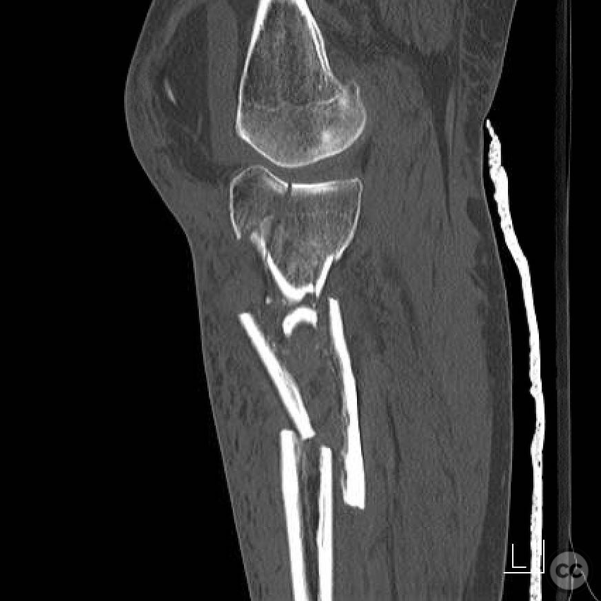
AO/OTA 41-C3 Tibial Plateau Fracture with Metaphyseal Comminution
Score and Comment on this Case
Clinical Details
Clinical and radiological findings: A 45-year-old male presented following a high-energy motor vehicle collision. The patient sustained an isolated closed injury to the proximal tibia with significant swelling and no neurovascular compromise. Radiographs and CT imaging revealed a complex comminuted fracture of the tibial plateau, classified as AO/OTA 41-C3, with extensive metaphyseal comminution. There was no evidence of associated ligamentous injury.
Preoperative Plan
Planning remarks: The preoperative plan involved a dual approach for addressing the articular and metaphyseal components. An anterolateral approach was planned for direct visualization and reduction of the articular surface, with the intention to use fragment-specific reduction techniques. For the metaphyseal comminution, a minimally invasive submuscular bridge plating technique was planned to provide relative stability.
Surgical Discussion
Patient positioning: The patient was positioned supine on a radiolucent table with a bump under the ipsilateral hip to allow for slight external rotation of the limb, facilitating access to both the anterolateral and posteromedial aspects of the proximal tibia.
Anatomical surgical approach: A longitudinal anterolateral incision was made, extending from Gerdy's tubercle distally along the tibial shaft. The iliotibial band was split in line with its fibers, and a submeniscal arthrotomy was performed to expose the lateral tibial plateau. A separate posteromedial incision was made to address any posterior fragments if necessary.
Operative remarks:The articular fragments were reduced using pointed reduction forceps and temporarily stabilized with K-wires. Interfragmentary compression was achieved using lag screws for absolute stability. A long 4.5mm locking compression plate was applied submuscularly along the lateral aspect of the tibia, bridging the metaphyseal comminution and providing relative stability. Care was taken to preserve soft tissue attachments and maintain fracture hematoma.
Postoperative protocol: Postoperatively, the patient was placed in a hinged knee brace allowing for immediate passive range of motion exercises. Weight-bearing was restricted to toe-touch for 6 weeks, progressing to partial weight-bearing at 8 weeks, and full weight-bearing as tolerated by 12 weeks.
Follow up: Not specified
Orthopaedic implants used: 4.5mm Synthes LCP locking plate, K-wires, lag screws
Search for Related Literature

orthopaedic_trauma
- United States , Seattle
- Area of Specialty - General Trauma
- Position - Specialist Consultant

Industry Sponsership
contact us for advertising opportunities
Article viewed 327 times
23 Jul 2025
Add to Bookmarks
Full Citation
Cite this article:
Surname, Initial. (2025). AO/OTA 41-C3 Tibial Plateau Fracture with Metaphyseal Comminution. Journal of Orthopaedic Surgery and Traumatology. Case Report 13489835 Published Online Jul 23 2025.