Complex Proximal Femur and Tibial Plateau Fracture with Soft Tissue Compromise
Score and Comment on this Case
Clinical Details
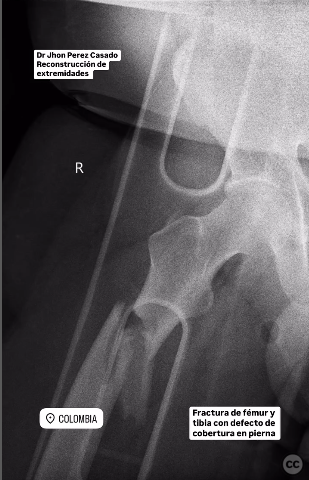
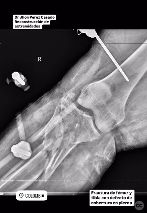
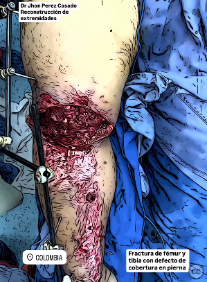
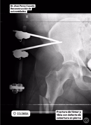
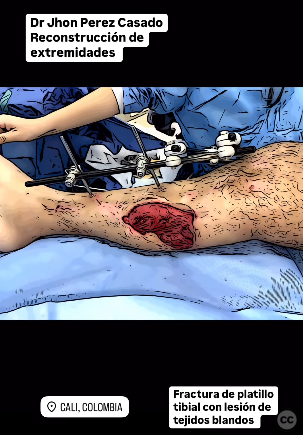
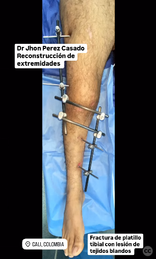
Clinical and radiological findings: The patient presented with a closed fracture of the proximal femur and a Gustilo-Anderson IIIC fracture of the tibia, indicating a severe open fracture with arterial injury requiring repair. Initial management included damage control measures with a trauma tutor and surgical washout. The tibial plateau fracture was in malunion due to complex soft tissue injury.
Preoperative Plan
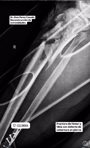
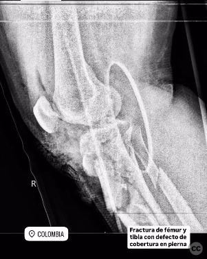
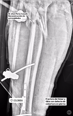
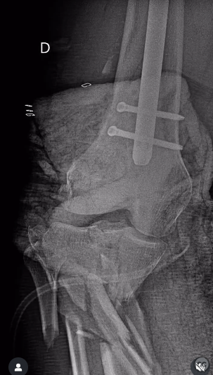
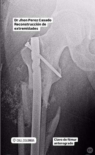
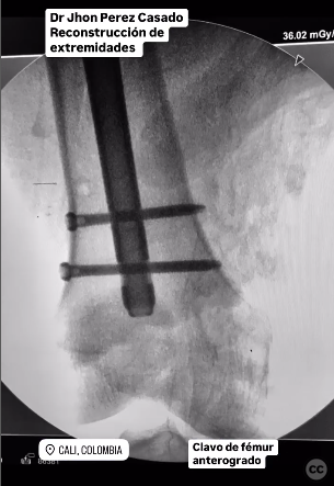
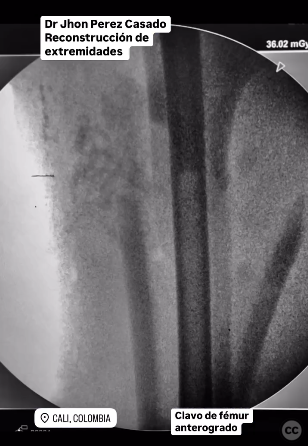
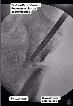
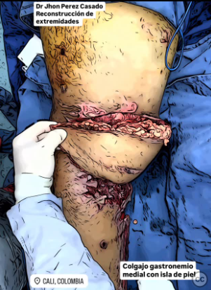
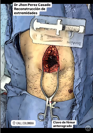
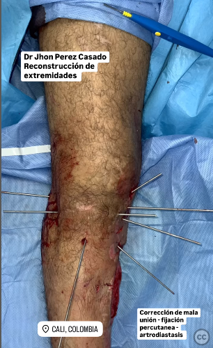
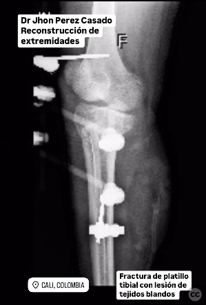
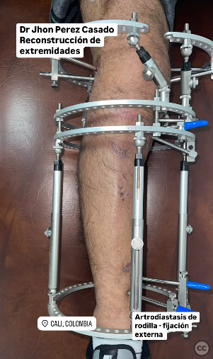
Planning remarks: The preoperative plan involved osteosynthesis of the femur using an anterograde intramedullary nail, application of bioactive crystal bone substitute, and negative pressure wound therapy for the leg. For the tibial plateau, removal of initial damage control scaffolding, soft tissue cleansing, closure flap, and percutaneous fixation were planned, followed by protection and arthrodiastasis using a circular external fixator.
Surgical Discussion
Patient positioning: The patient was positioned supine for the femoral procedure and likely maintained in a similar position for subsequent tibial interventions to facilitate access to both the proximal femur and tibia.
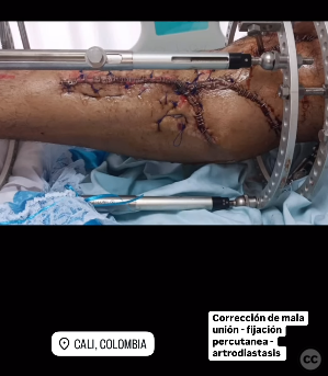
Anatomical surgical approach: A longitudinal incision was made for the femoral approach to accommodate the insertion of an anterograde intramedullary nail. For the tibial plateau, a percutaneous approach was utilized for fixation, with additional soft tissue management through flap closure techniques.
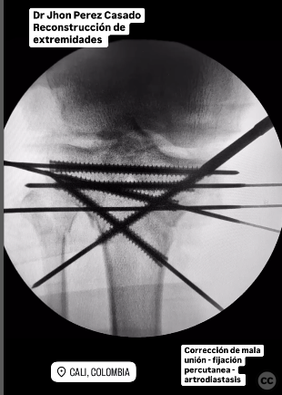
Operative remarks:The surgeon noted the complexity of managing the malunion and soft tissue injury. The use of bioactive crystal bone substitute was emphasized for both femoral and tibial reconstructions. The application of a circular external fixator provided necessary stability and facilitated soft tissue healing through arthrodiastasis.
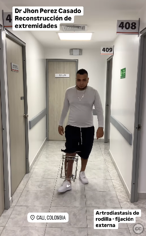
Postoperative protocol: Postoperative rehabilitation included walking training and muscle strengthening exercises. The use of an external fixator allowed for early mobilization while protecting the surgical site.
Follow up: Not specified.
Orthopaedic implants used: Anterograde intramedullary nail, bioactive crystal bone substitute, circular external fixator.
Search for Related Literature
Jhon Perez Casado
- Colombia , Cali - Valle del Cauca
- Area of Specialty - Lower Limb
- Position - Specialist Consultant

Industry Sponsership
contact us for advertising opportunities
Article viewed 307 times
25 Jul 2025
Add to Bookmarks
Full Citation
Cite this article:
Perez Casado, J.J.. (2025). Complex Proximal Femur and Tibial Plateau Fracture with Soft Tissue Compromise. Journal of Orthopaedic Surgery and Traumatology. Case Report 11993735 Published Online Jul 25 2025.