Combined Anterior Column and Posterior Hemi-Transverse Acetabular Fracture: Ilioinguinal Approach with Focal Plate Fixation
Score and Comment on this Case
Clinical Details
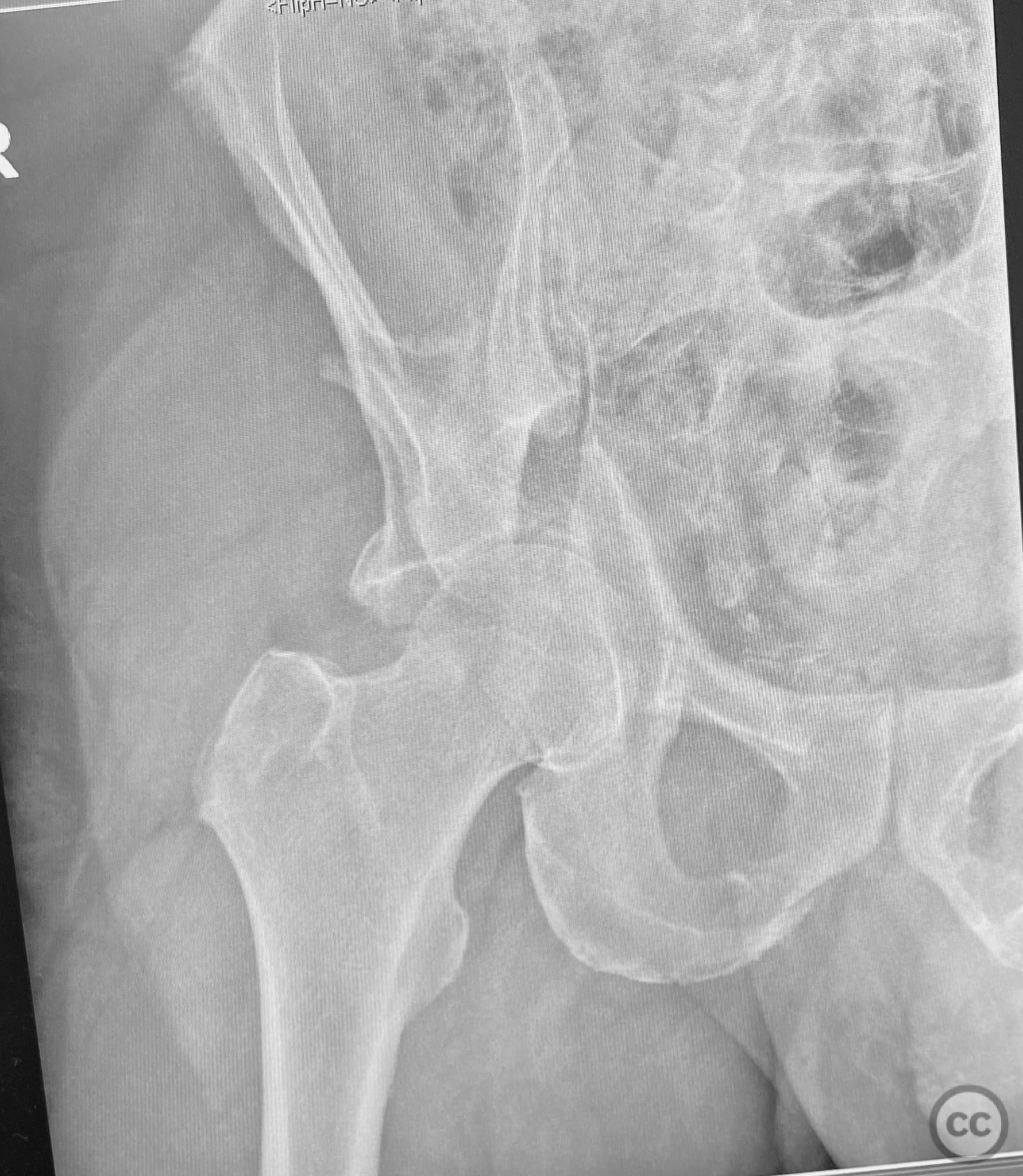
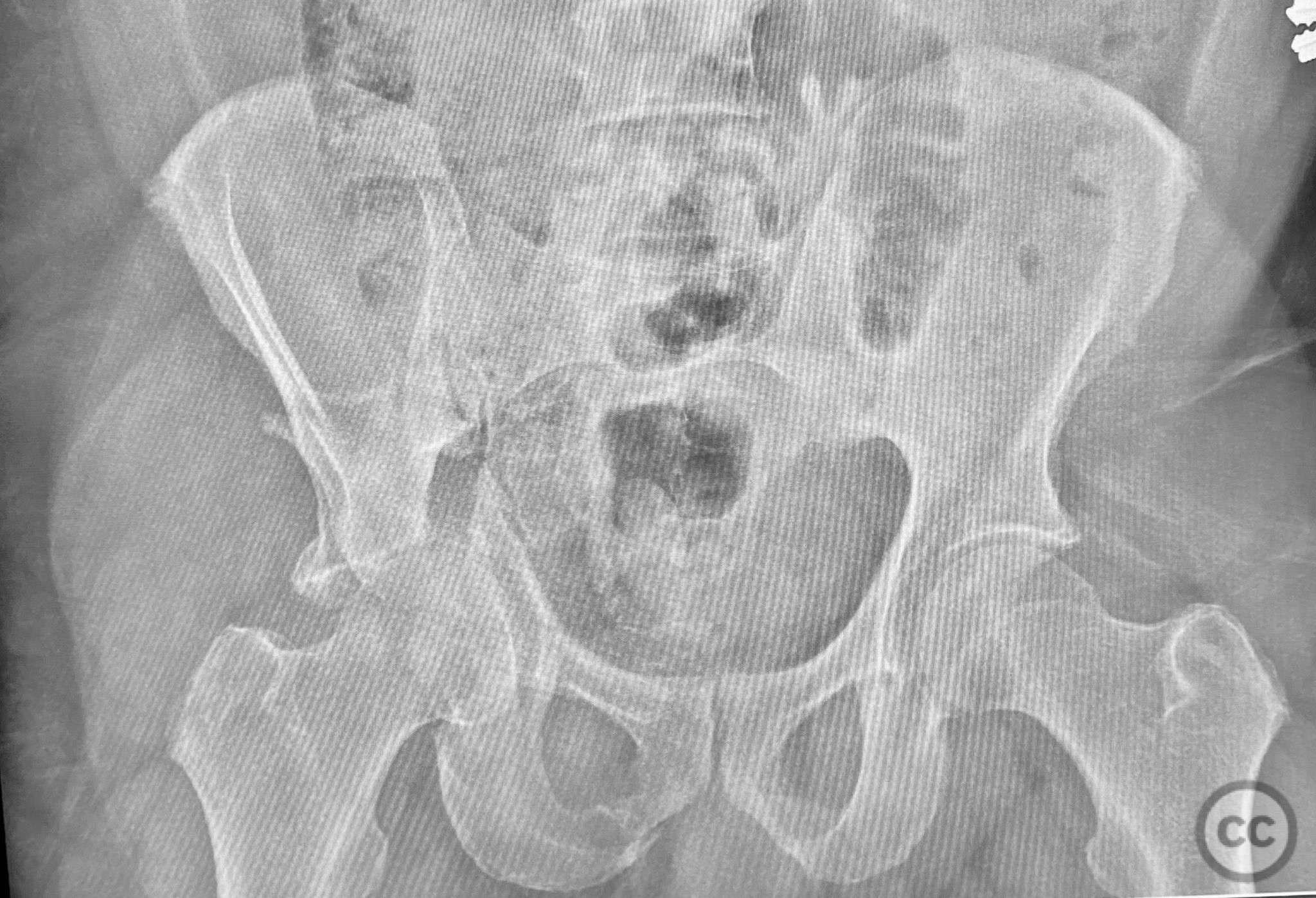
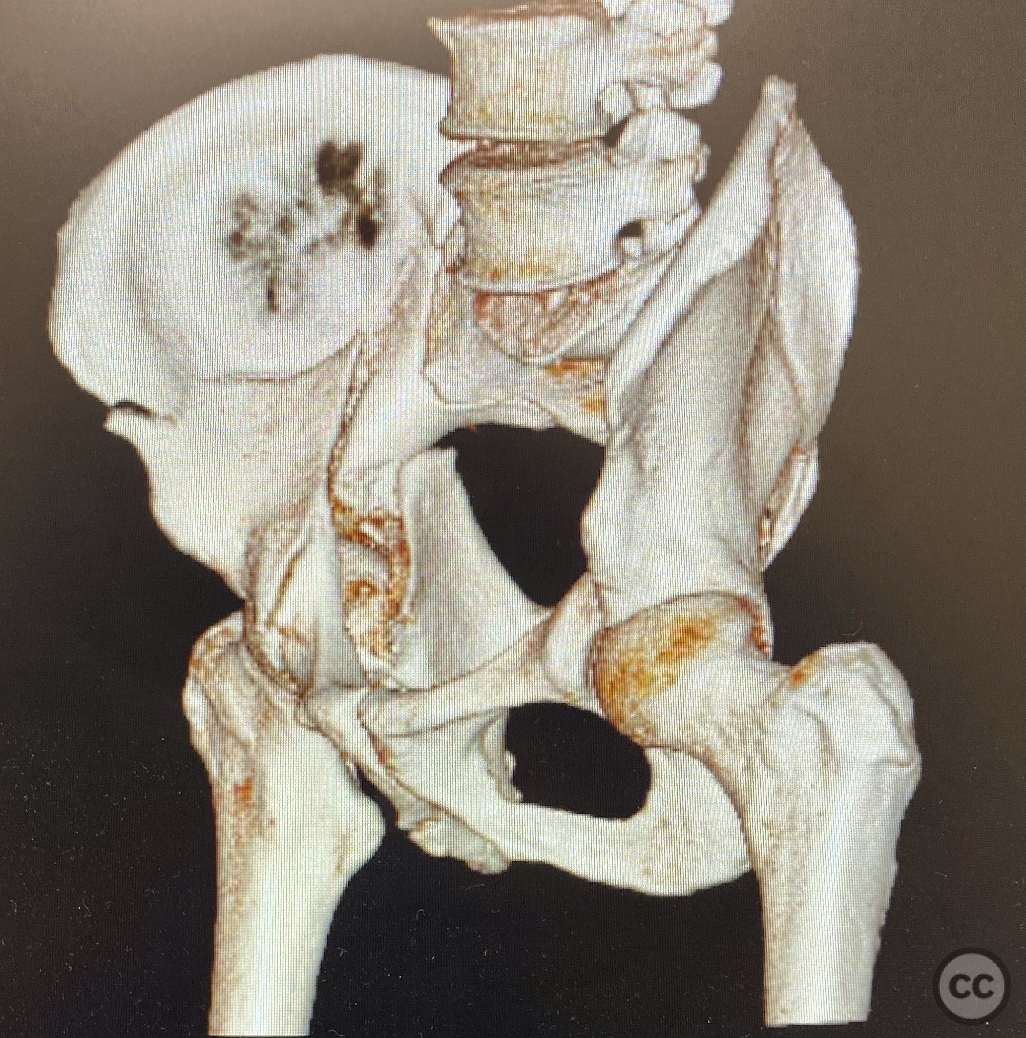
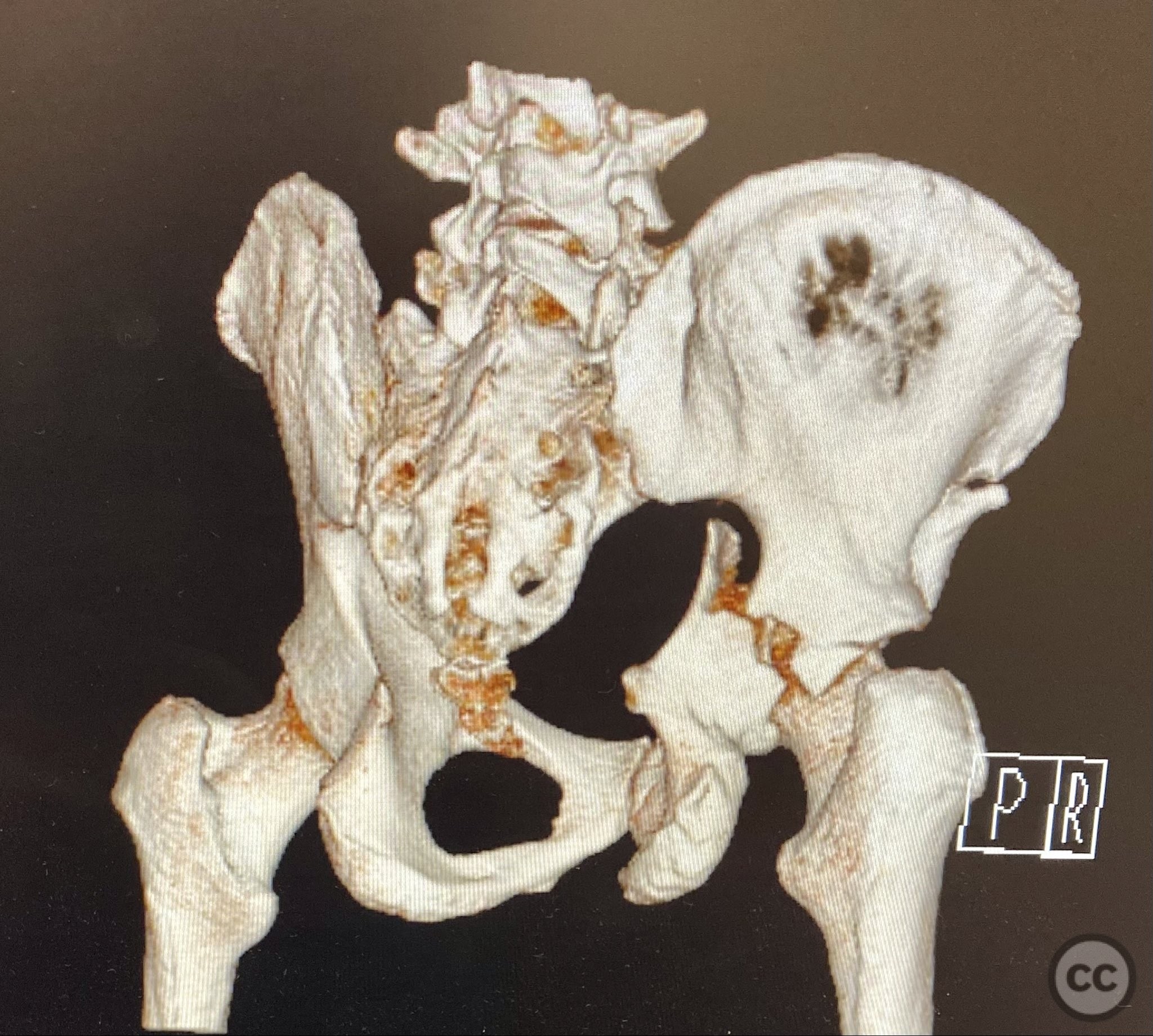
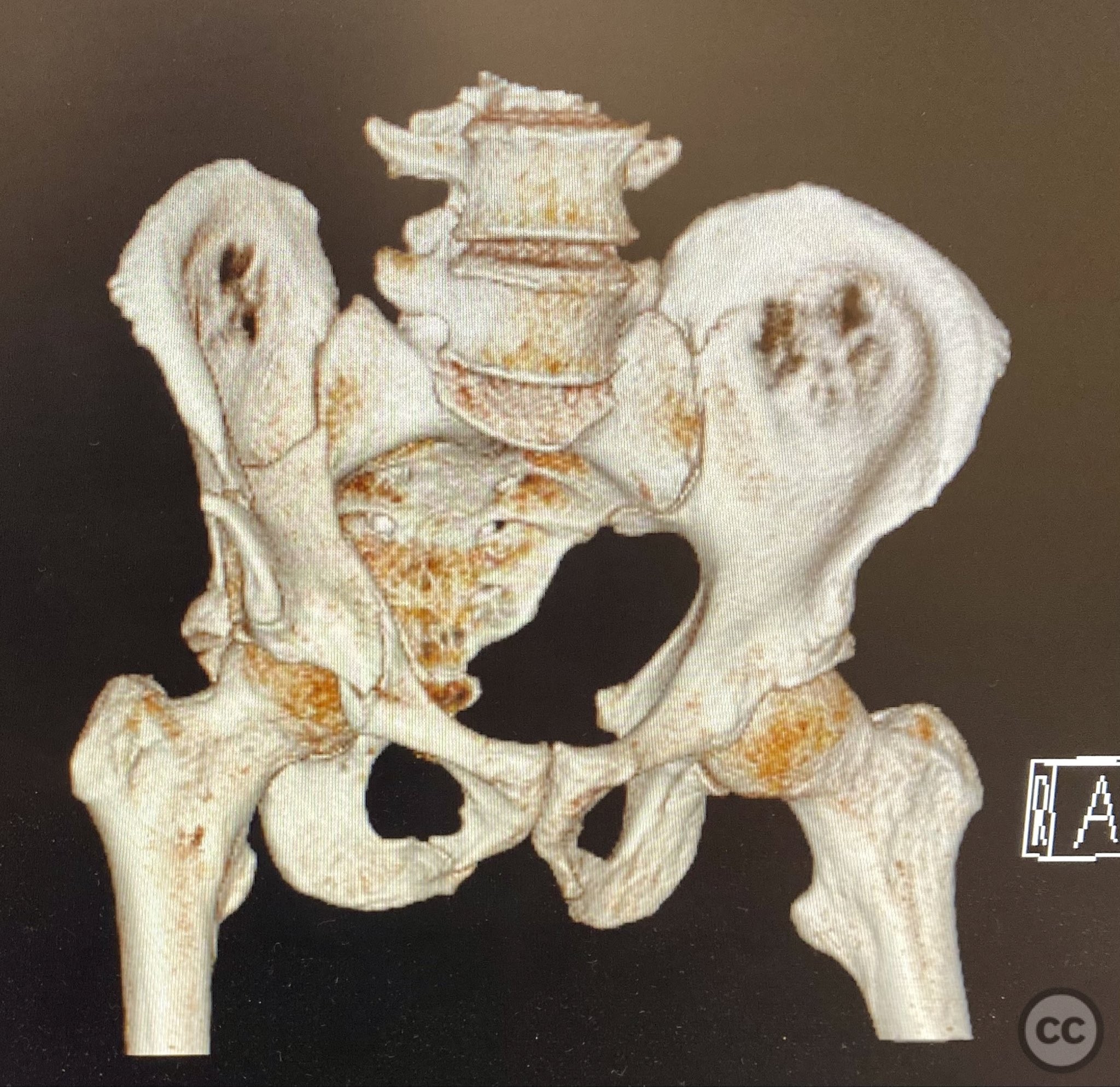
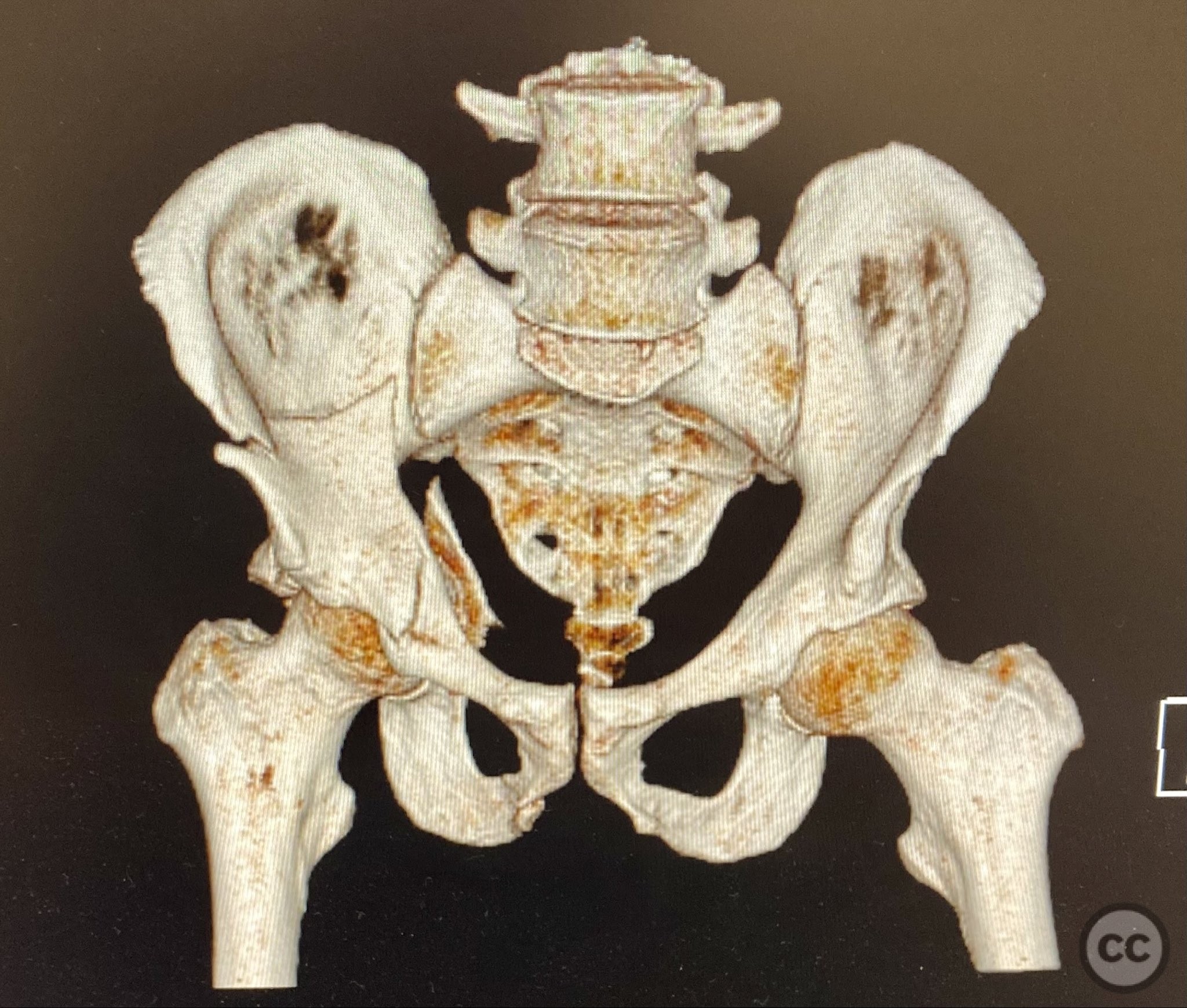
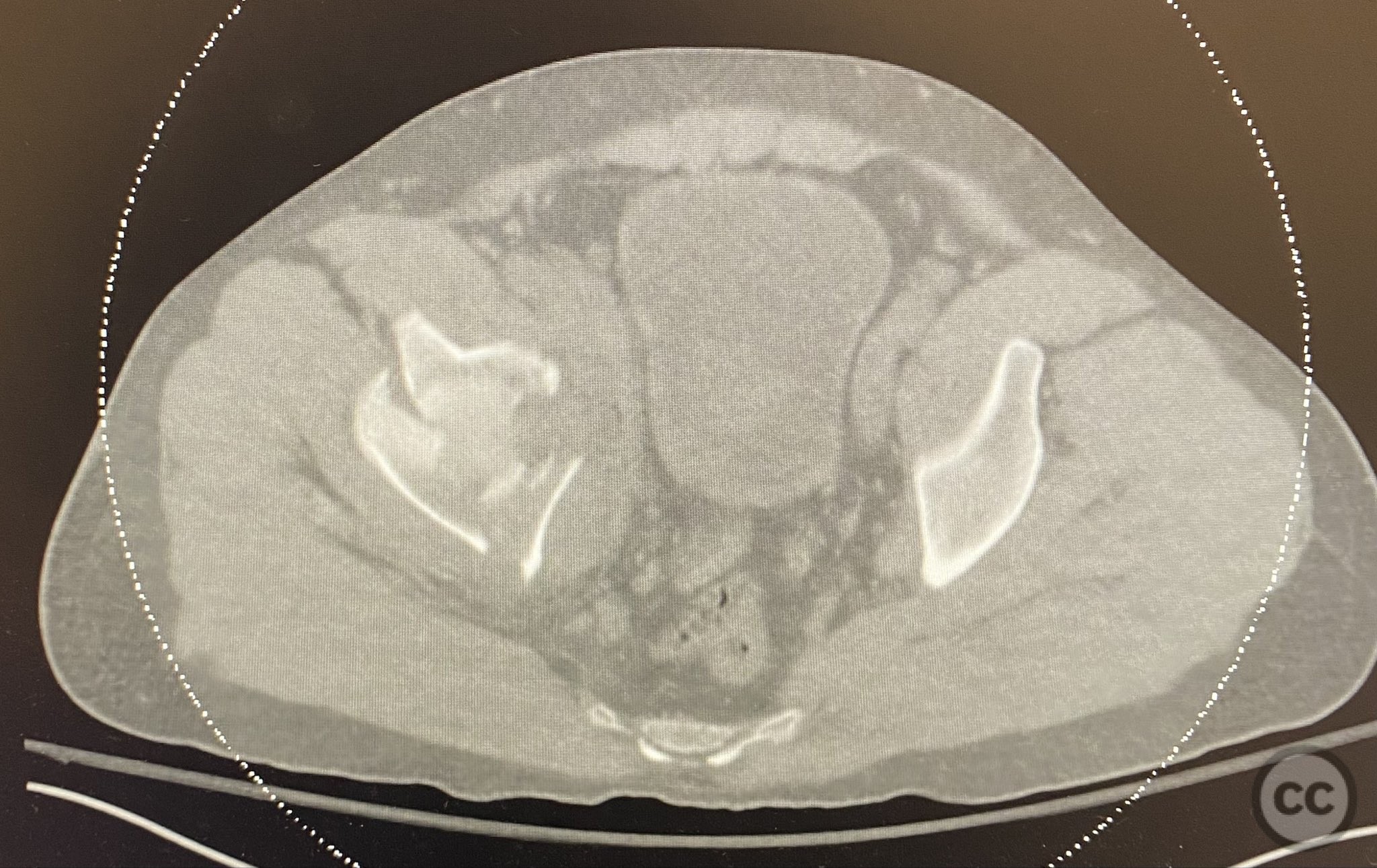
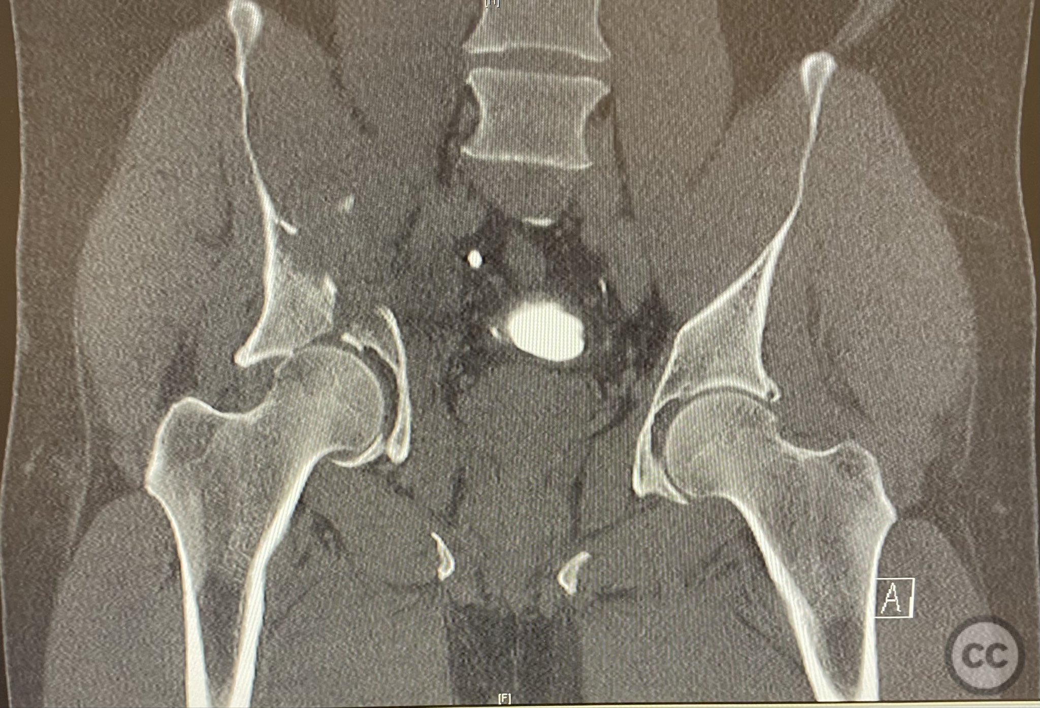
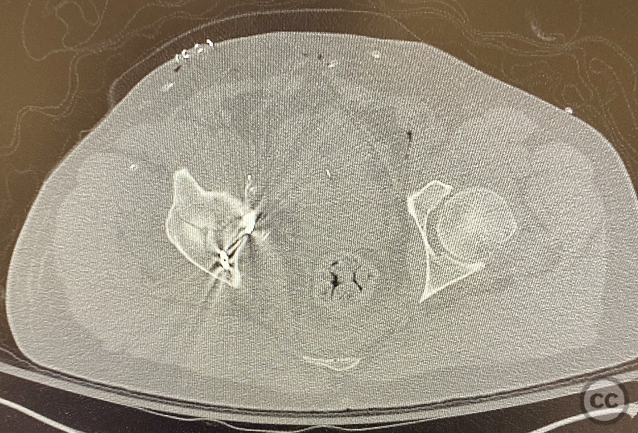
Clinical and radiological findings: A patient presented with a combined anterior column and posterior hemi-transverse (AC/PHTr) acetabular fracture. Plain radiographs demonstrated multiple displaced fragments involving the acetabular dome and femoral head region, with clear disruption of the anterior column and posterior transverse components. Advanced imaging, including axial and coronal CT, provided detailed visualization of cortical surface displacement, fragment orientation, and associated soft tissue status. The edge of the intact dome and femoral head injuries were identified, as well as a residual defect at the edge of the dome following reduction. AO/OTA classification: 62B2 (anterior column with posterior hemitransverse).
Preoperative Plan
Planning remarks: The preoperative plan was to utilize an ilioinguinal approach, specifically employing the iliac, middle, and intrapelvic intervals for exposure. The plan included anatomical reduction of the anterior column and posterior hemi-transverse fragments using bone clamps, followed by stabilization with buttressing plates and interfragmentary lag screws. No intraoperative traction was planned.
Surgical Discussion
Patient positioning: The patient was positioned supine on a radiolucent table, with no traction applied.
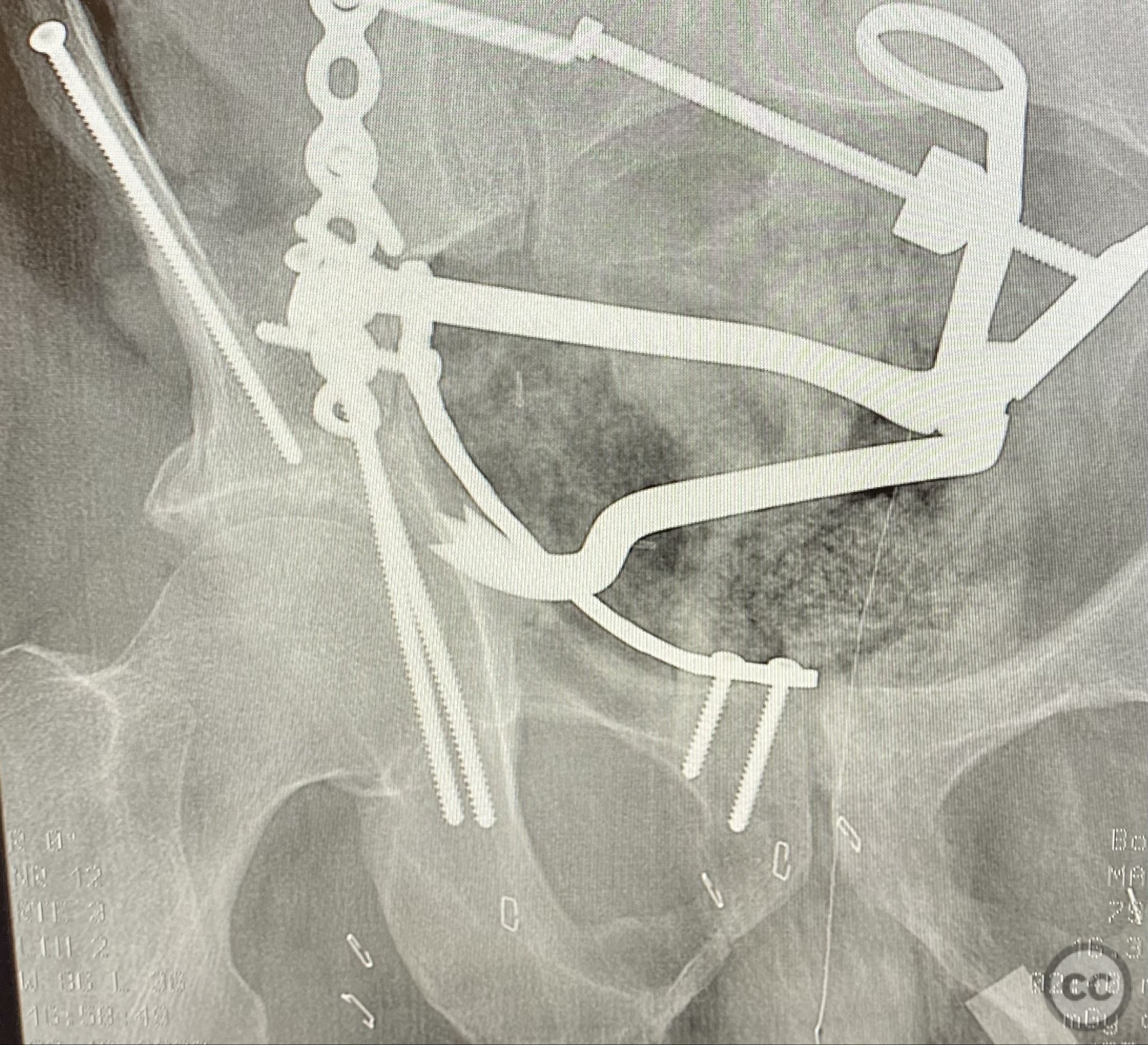
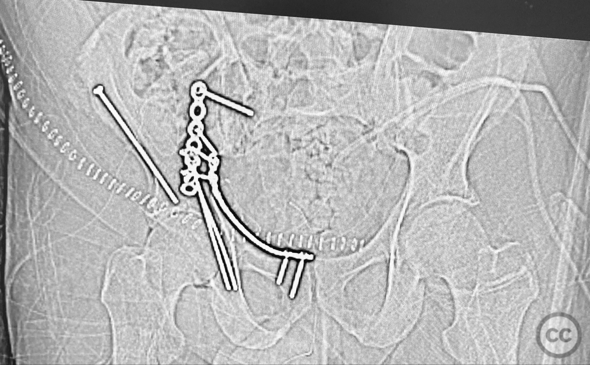
Anatomical surgical approach: A standard ilioinguinal approach was performed, sequentially developing the iliac, middle, and intrapelvic windows. Subperiosteal dissection exposed the fractured anterior column and quadrilateral plate. The fracture planes were meticulously debrided of hematoma and interposed tissue. Bone clamps were applied to achieve reduction of the anterior column and posterior hemi-transverse fragments. Buttressing plates were contoured and applied to the pelvic brim and quadrilateral surface, with interfragmentary lag screws placed through the plates to secure the reduction while maintaining clamp control.
Operative remarks:Surface renderings from preoperative imaging facilitated intraoperative correlation between radiographic and fluoroscopic views, aiding in precise interpretation of fragment orientation during reduction. The use of focal plates allowed for maintenance of provisional reductions with bone holding clamps during definitive fixation. Restoration of radiographic lines was confirmed on intraoperative AP fluoroscopy, while postoperative axial CT demonstrated successful central dome repair with a residual edge crush defect at the dome margin.
Postoperative protocol: Postoperatively, early passive range of motion exercises were initiated with toe-touch weight bearing for 8 weeks. Progressive weight bearing was advanced according to radiographic evidence of healing.
Follow up: Not specified
Orthopaedic implants used: Focal buttressing plates, interfragmentary lag screws
Search for Related Literature


Industry Sponsership
contact us for advertising opportunities
Article viewed 680 times
12 Sep 2025
Add to Bookmarks
Full Citation
Cite this article:
Routt, ML. (2025). Combined Anterior Column and Posterior Hemi-Transverse Acetabular Fracture: Ilioinguinal Approach with Focal Plate Fixation. Journal of Orthopaedic Surgery and Traumatology. Case Report 11914927 Published Online Sep 12 2025.