 went thru his boot then thru and thru his foot between the firs_2.jpg)
 went thru his boot then thru and thru his foot between the first(.jpg)
 went thru his boot then thru and thru his foot between the firs_1.jpg)
 after a _motocross mishap._._._._He has a combine_3.jpg)
 after a _motocross mishap._._._._He has a combine_5.jpg)
 after a _motocross mishap._._._._He has a combine_4.jpg)
 after a _motocross mishap._._._._He has a combine_7.jpg)
 after a _motocross mishap._._._._He has a combine_6.jpg)
 after a _motocross mishap._._._._He has a combine_8.jpg)
 after a _motocross mishap._._._._He has a combine_9.jpg)
 after a _motocross mishap._._._._He has a combined(.jpg)
 after a _motocross mishap._._._._He has a combine_1.jpg)
 after a _motocross mishap._._._._He has a combine_2.jpg)
Complex Lisfranc and Lateral Column Crush Injury with Open Plantar and Dorsal Wounds.
Score and Comment on this Case
Clinical Details
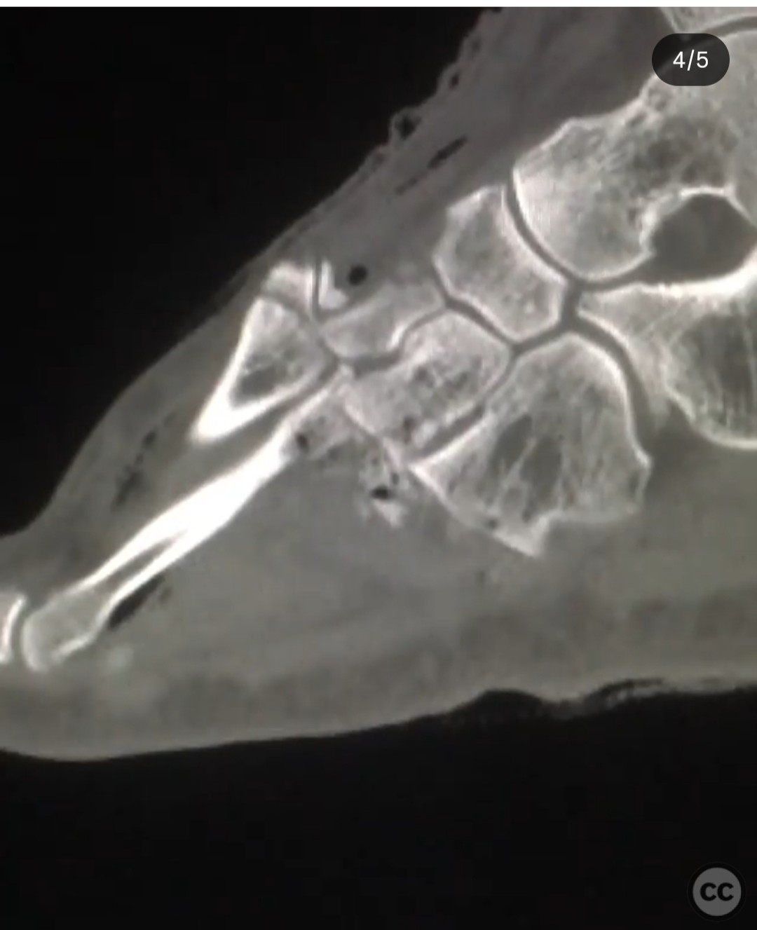
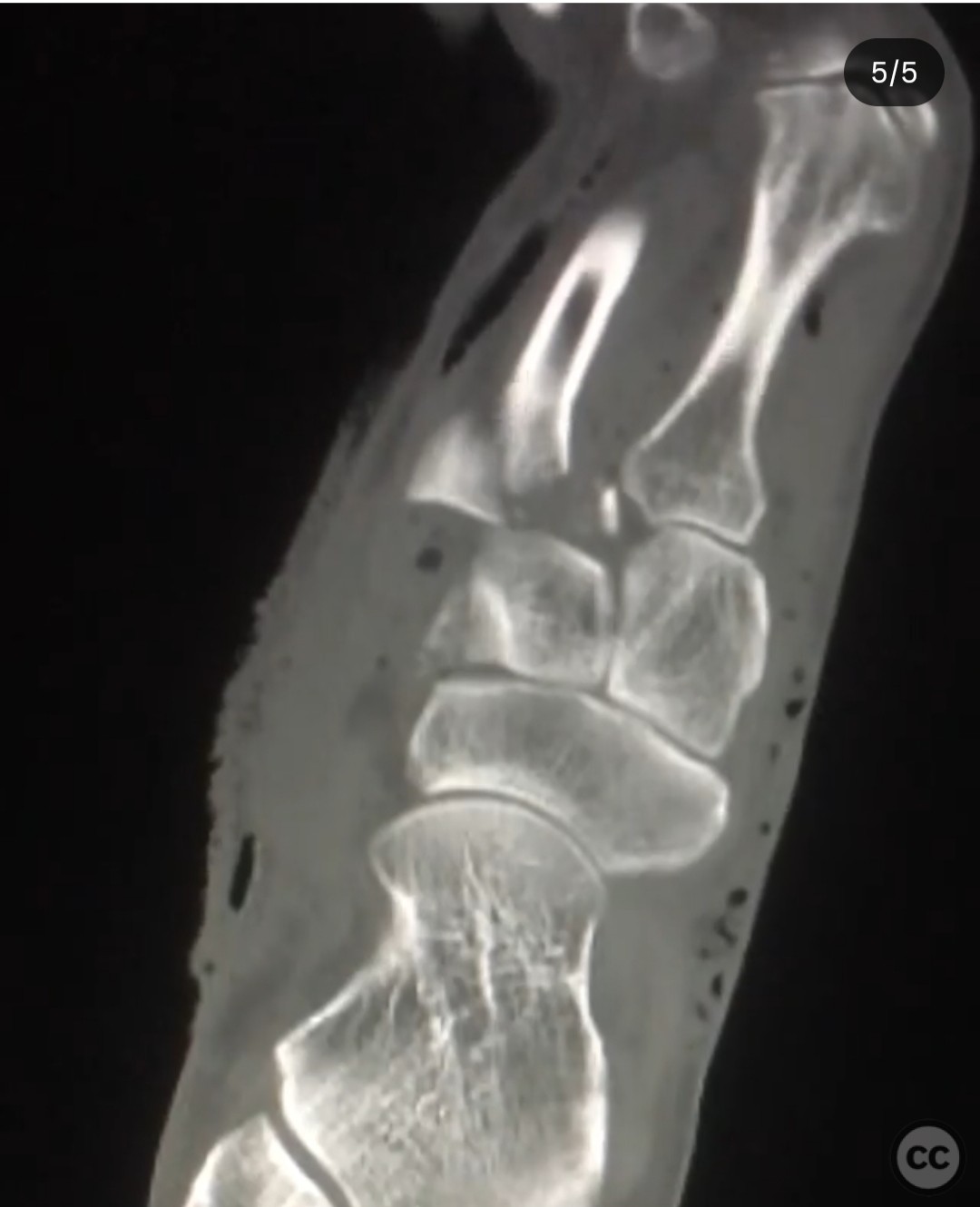
Clinical and radiological findings: A 31-year-old male sustained a high-energy open foot injury following a motocross accident, characterized by a through-and-through wound between the first and second ray, with both dorsal and plantar involvement (Gustilo-Anderson Type 3A). Radiological assessment revealed a combined Lisfranc injury involving tarso-metatarsal fracture dislocations of the second to fifth rays, inter-cuneiform instability, and a lateral column crush injury, including a cuboid fracture with calcaneo-cuboid subluxation. The lateral column was notably shortened, indicating a nutcracker-type injury.
Preoperative Plan
Planning remarks: The preoperative plan involved a staged approach, initially addressing the medial column by stabilizing the inter-cuneiform joint disruption, followed by reduction of the second metatarsal to its keystone position. Reconstruction of the third metatarsal to its corresponding cuneiform was planned, with attention to the intermetatarsal ligaments for the fourth and fifth metatarsals. The lateral column was to be managed with a spanning plate to maintain length due to the inadequacy of external fixation in this pattern.
Surgical Discussion
Patient positioning: Supine positioning on the operating table with the injured foot elevated and prepared for sterile field access.
Anatomical surgical approach: A dual approach was utilized: a dorsal incision for direct access to the Lisfranc complex and inter-cuneiform space, and a lateral incision to address the lateral column crush injury. The dorsal approach allowed for visualization and stabilization of the inter-cuneiform instability and tarso-metatarsal dislocations, while the lateral approach facilitated reduction and fixation of the cuboid fracture and calcaneo-cuboid subluxation.
Operative remarks:The surgeon noted the importance of addressing both medial and lateral column injuries to prevent future deformities. The medial column was rigidly fixed to ensure stability, while the lateral column required flexible fixation due to the complex nature of the injury. The use of a spanning plate effectively maintained lateral column length without relying on ligamentotaxis, which was compromised in this case.
Postoperative protocol: Postoperative rehabilitation included non-weight bearing on the affected limb for six weeks, followed by progressive weight-bearing as tolerated. Physical therapy focused on range of motion exercises and gradual strengthening.
Follow up: Not specified.
Orthopaedic implants used: Spanning plate for lateral column stabilization, screws for rigid fixation of the medial column, Kirschner wires for temporary stabilization.
Search for Related Literature

orthopaedic_trauma
- United States , Seattle
- Area of Specialty - General Trauma
- Position - Specialist Consultant

Industry Sponsership
contact us for advertising opportunities
Article viewed 314 times
23 Jul 2025
Add to Bookmarks
Full Citation
Cite this article:
Surname, Initial. (2025). Complex Lisfranc and Lateral Column Crush Injury with Open Plantar and Dorsal Wounds.. Journal of Orthopaedic Surgery and Traumatology. Case Report 11522699 Published Online Jul 23 2025.